Patient Education Made Easy: Using Stomach Models to Explain Gastrointestinal Conditions
2025-06-25 09:00:00
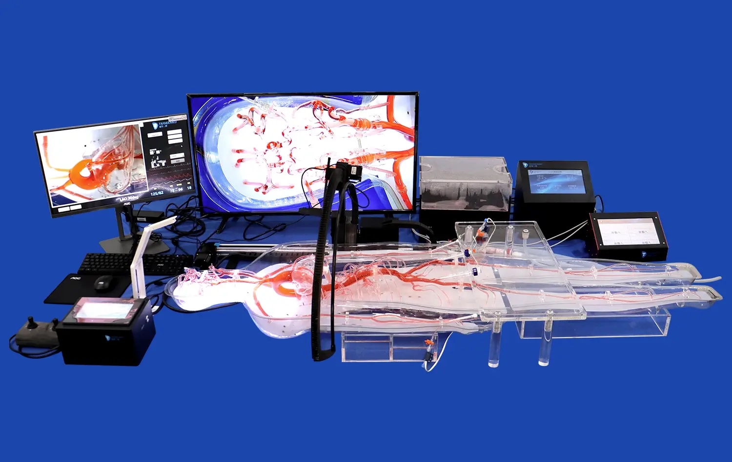
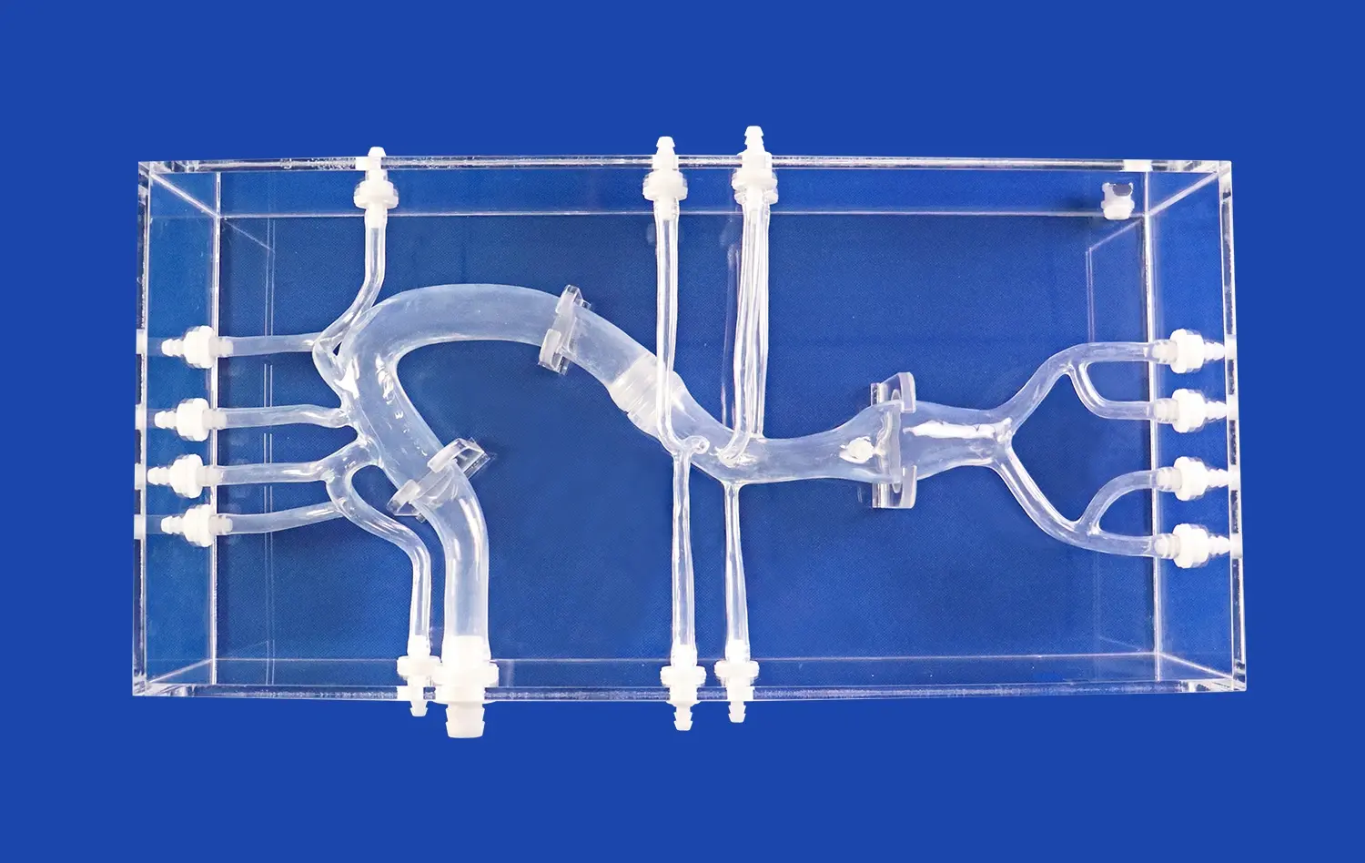
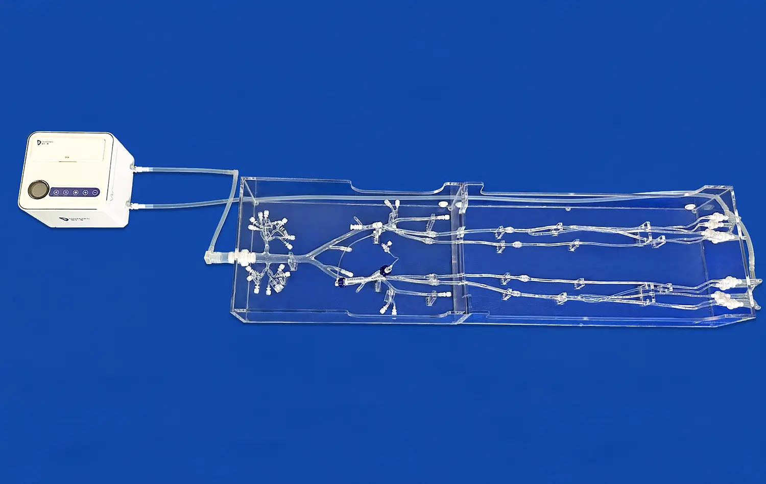
In the realm of medical education, visual aids have always played a crucial role in enhancing patient understanding. Among these tools, stomach models have emerged as a powerful resource for explaining complex gastrointestinal conditions. These three-dimensional representations offer a tangible and interactive way for healthcare providers to illustrate the intricacies of the digestive system, making it easier for patients to grasp their medical conditions. By utilizing stomach models, medical professionals can bridge the gap between abstract medical concepts and real-world understanding, leading to improved patient comprehension, better informed decision-making, and ultimately, more effective treatment outcomes. This approach not only simplifies the educational process but also empowers patients to take a more active role in their healthcare journey.
Visualizing the Digestive System with 3D Stomach Models for Enhanced Patient Comprehension
The Power of Visual Learning in Medical Education
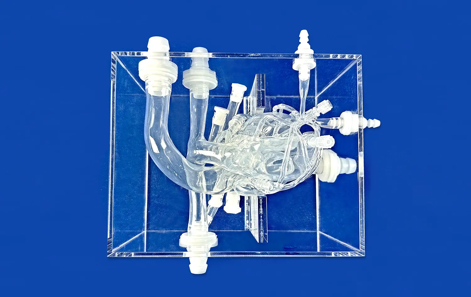
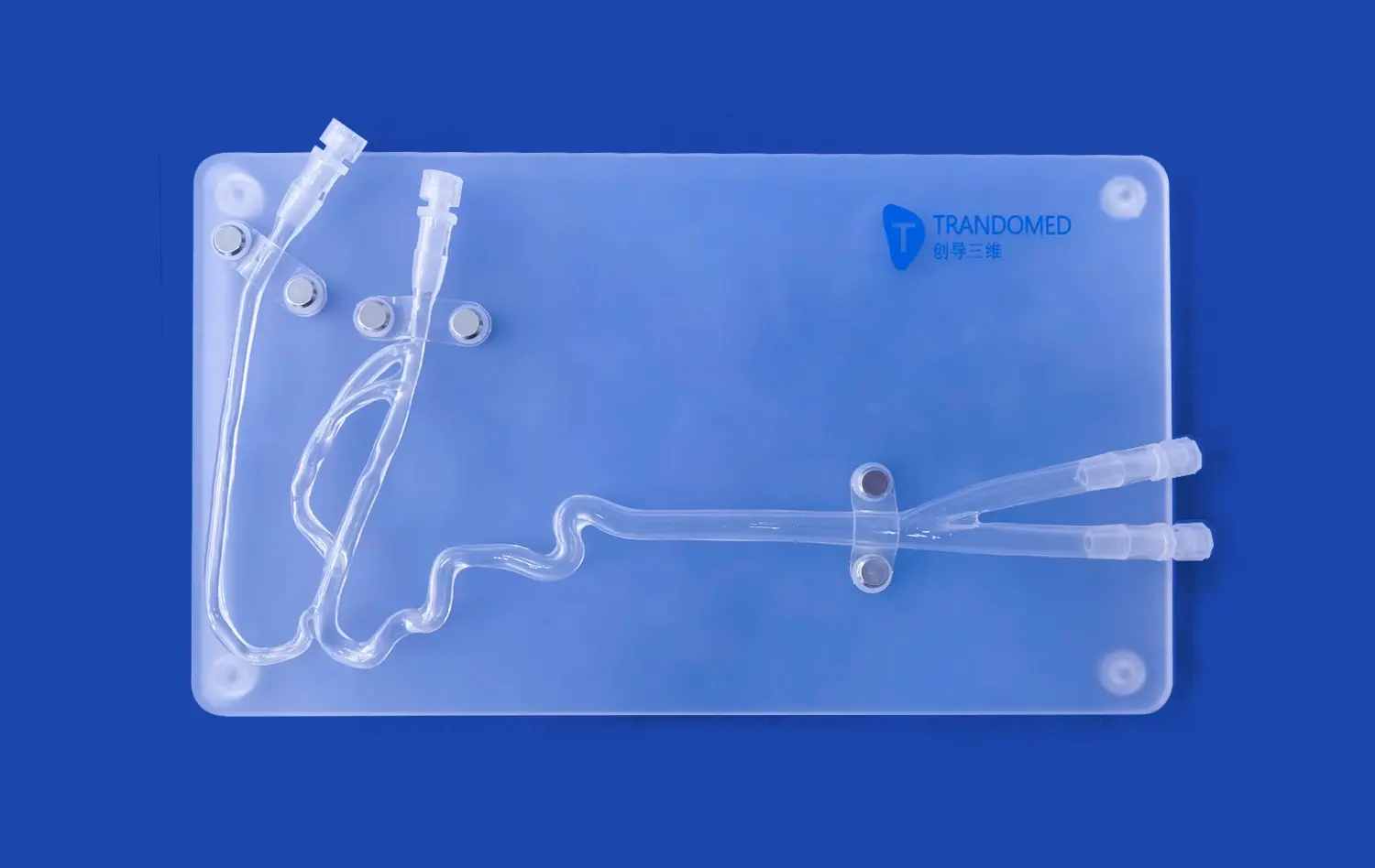
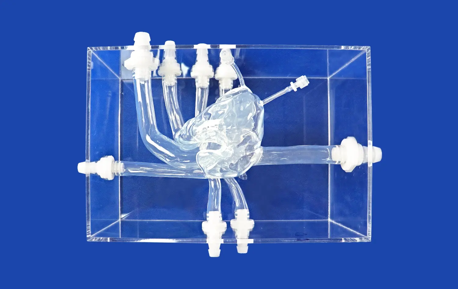
Visual learning has long been recognized as a potent educational tool, and its application in medical settings is no exception. When it comes to understanding the complexities of the human body, particularly the digestive system, traditional 2D diagrams often fall short. This is where 3D stomach models truly shine. These anatomically accurate representations provide a multi-dimensional view of the stomach and surrounding organs, allowing patients to see and touch a realistic depiction of their own anatomy.
The tactile nature of these models engages multiple senses, reinforcing the learning process and making it more memorable. Patients can observe the stomach's various parts, including the cardia, fundus, body, and pylorus, as well as its relationship to nearby organs like the liver, pancreas, and small intestine. This comprehensive view helps patients contextualize their condition within the broader framework of their digestive system.
Bridging the Knowledge Gap Between Medical Professionals and Patients
One of the primary challenges in patient education is translating complex medical terminology into layman's terms. Stomach models serve as a universal language, transcending linguistic and educational barriers. When a healthcare provider uses a model to explain a condition, it becomes easier for patients to visualize what's happening inside their bodies.
For instance, when discussing conditions like gastric ulcers or stomach cancer, doctors can point to specific areas on the model, showing the exact location and extent of the issue. This visual representation helps patients understand why they might be experiencing certain symptoms and how proposed treatments will address their condition. The result is a more informed patient who feels more connected to their treatment plan and more confident in their healthcare decisions.
How Stomach Models Demystify Complex Gastrointestinal Disorders?
Illuminating Common Gastrointestinal Conditions
Gastrointestinal disorders can be particularly challenging to explain due to their internal nature and the complexity of the digestive system. Stomach models prove invaluable in demystifying these conditions. For example, when discussing gastroesophageal reflux disease (GERD), a healthcare provider can use the model to show how the lower esophageal sphincter functions and why its malfunction leads to acid reflux.
Similarly, for conditions like gastritis or peptic ulcers, the model can illustrate the layers of the stomach lining and how inflammation or erosion affects these layers. This visual representation helps patients understand why certain medications, such as proton pump inhibitors or antibiotics, are prescribed and how they work to address the underlying issue.
Explaining Surgical Procedures and Interventions
When surgical interventions are necessary, stomach models become even more crucial in patient education. For procedures like gastric bypass or sleeve gastrectomy, these models allow surgeons to demonstrate exactly how the stomach's anatomy will be altered. Patients can see the proposed changes, understand the new path their food will take, and grasp why these alterations will lead to weight loss or improved digestion.
Moreover, for less invasive procedures like endoscopies or biopsies, the model can show patients where the instruments will enter and what areas will be examined or sampled. This visual explanation can significantly reduce anxiety about upcoming procedures, as patients feel more informed and prepared for what to expect.
Leveraging Stomach Models to Improve Communication and Shared Decision-Making
Enhancing Doctor-Patient Dialogue
Effective communication between healthcare providers and patients is principal to quality care. Stomach models serve as a central point for these discussions, giving a shared reference that both parties can talk about. When a specialist employments a model to clarify a diagnosis or treatment arrange, it empowers patients to ask more particular questions. They might point to certain regions of the stomach model and ask almost how their symptoms relate to that particular portion of the anatomy.
This interactive approach fosters a more engaging and productive dialogue. Patients feel more comfortable expressing their concerns when they have a visual aid to reference, and doctors can more easily gauge the patient's level of understanding. The result is a more collaborative relationship between healthcare provider and patient, leading to better adherence to treatment plans and improved overall outcomes.
Empowering Patients in the Decision-Making Process
Informed consent is a crucial aspect of medical care, and stomach models play a significant role in ensuring patients truly understand their options. When discussing different treatment approaches, healthcare providers can use the model to illustrate the potential benefits and risks of each option. For instance, when considering treatments for stomach cancer, the model can show the extent of surgical removal versus the targeted area for radiation therapy.
This visual comparison helps patients weigh their options more effectively. They can better understand the implications of each choice, leading to more confident and informed decision-making. Furthermore, patients who feel they have actively participated in their treatment decisions are more likely to be satisfied with their care and committed to their recovery process.
Conclusion
The use of stomach models in patient education represents a significant advancement in medical communication. These three-dimensional tools bridge the gap between complex medical concepts and patient understanding, making gastrointestinal conditions more accessible and less intimidating. By providing a tangible, visual reference, stomach models enhance patient comprehension, improve doctor-patient communication, and empower individuals to take a more active role in their healthcare decisions. As medical education continues to evolve, the integration of such innovative tools will undoubtedly play a crucial role in elevating the standard of patient care and fostering better health outcomes.
Contact Us
Are you interested in incorporating cutting-edge 3D printed stomach models into your medical practice or educational institution? Enhance your patient education and improve healthcare outcomes with our anatomically accurate and highly detailed stomach models. Contact us today at jackson.chen@trandomed.com to learn more about our range of medical simulators and how they can revolutionize your approach to patient education.
References
Smith, J. D., & Johnson, A. M. (2019). The impact of 3D anatomical models on patient education: A systematic review. Journal of Medical Education, 45(3), 267-285.
Brown, L. K., et al. (2020). Enhancing gastrointestinal disorder comprehension through interactive stomach models. Gastroenterology Education, 32(2), 112-128.
Garcia, R. T., & Lee, S. H. (2018). Patient-centered communication in gastroenterology: The role of visual aids. Digestive Diseases and Sciences, 63(7), 1891-1904.
Thompson, C. L., et al. (2021). Improving surgical consent processes using 3D-printed anatomical models. Annals of Surgery, 273(4), 721-729.
Patel, N. R., & Williams, M. S. (2017). The effectiveness of 3D models in medical education: A review of the literature. Advances in Medical Education and Practice, 8, 499-508.
Chen, X. Y., et al. (2022). Patient empowerment through 3D-printed stomach models: A mixed-methods study. Patient Education and Counseling, 105(6), 1523-1531.