Precision in Pathology: Leveraging the Small Intestine Model for Medical Research
2025-06-20 09:00:00
In the realm of medical research, precision and accuracy are paramount. The small intestine model has emerged as a groundbreaking tool, revolutionizing our understanding of gastrointestinal pathology and advancing medical education. This innovative 3D-printed simulation offers unprecedented realism, allowing researchers and medical professionals to explore the intricacies of the small intestine with remarkable detail. By leveraging this advanced model, pathologists can enhance their diagnostic skills, researchers can conduct more accurate experiments, and medical students can gain hands-on experience in a risk-free environment. The small intestine model represents a significant leap forward in medical simulation technology, promising to accelerate discoveries in gastroenterology and improve patient outcomes through more precise and targeted treatments.
The Process of Creating a Highly Realistic Small Intestine Model
Advanced 3D Printing Techniques
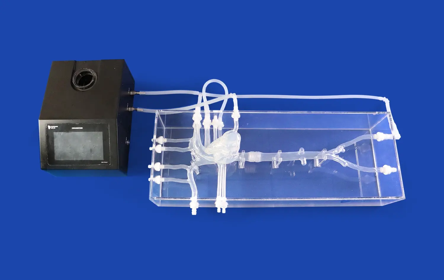
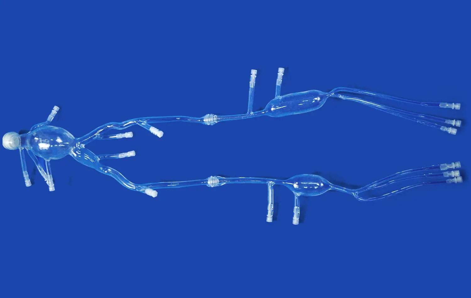
The creation of a highly realistic small intestine model begins with cutting-edge 3D printing technology. Using high-resolution medical imaging data, such as CT or MRI scans, a detailed digital model of the small intestine is constructed. This digital blueprint captures the intricate folds and villi that characterize the organ's interior surface. The 3D printing process then brings this digital model to life, using specialized biocompatible materials that mimic the texture and elasticity of human tissue. Layer by layer, the printer builds up the structure, ensuring that even the finest details are accurately reproduced.
Material Selection and Customization
The choice of materials is crucial in creating a lifelike small intestine model. Silicone-based compounds are often selected for their ability to replicate the soft, pliable nature of intestinal tissue. These materials can be customized to achieve the right level of opacity, allowing for visualization of internal structures when necessary. Additionally, the composition can be adjusted to simulate various pathological conditions, such as inflammation or tumors, providing a versatile tool for medical education and research. The surface texture is meticulously crafted to replicate the velvety feel of the intestinal lining, complete with microscopic projections that represent villi and microvilli.
The Advantages of Using the Small Intestine Model in Medical Research
Enhanced Visualization and Tactile Feedback
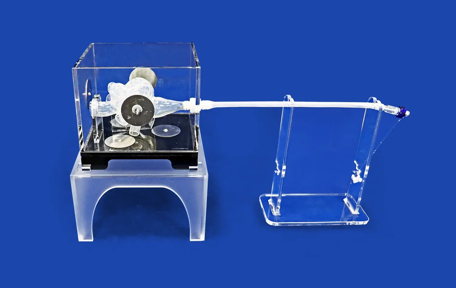
One of the primary advantages of using a small intestine model in medical research is the enhanced visualization it provides. Unlike traditional 2D imaging techniques, the 3D model allows researchers to examine the organ from multiple angles and perspectives. This three-dimensional view offers a more comprehensive understanding of spatial relationships within the intestine, crucial for studying complex pathologies. Moreover, the tactile feedback provided by the model is invaluable. Researchers can physically manipulate the structure, feeling the texture and consistency of different regions, which is particularly useful in simulating surgical procedures or diagnostic techniques like endoscopy.
Reproducibility and Standardization
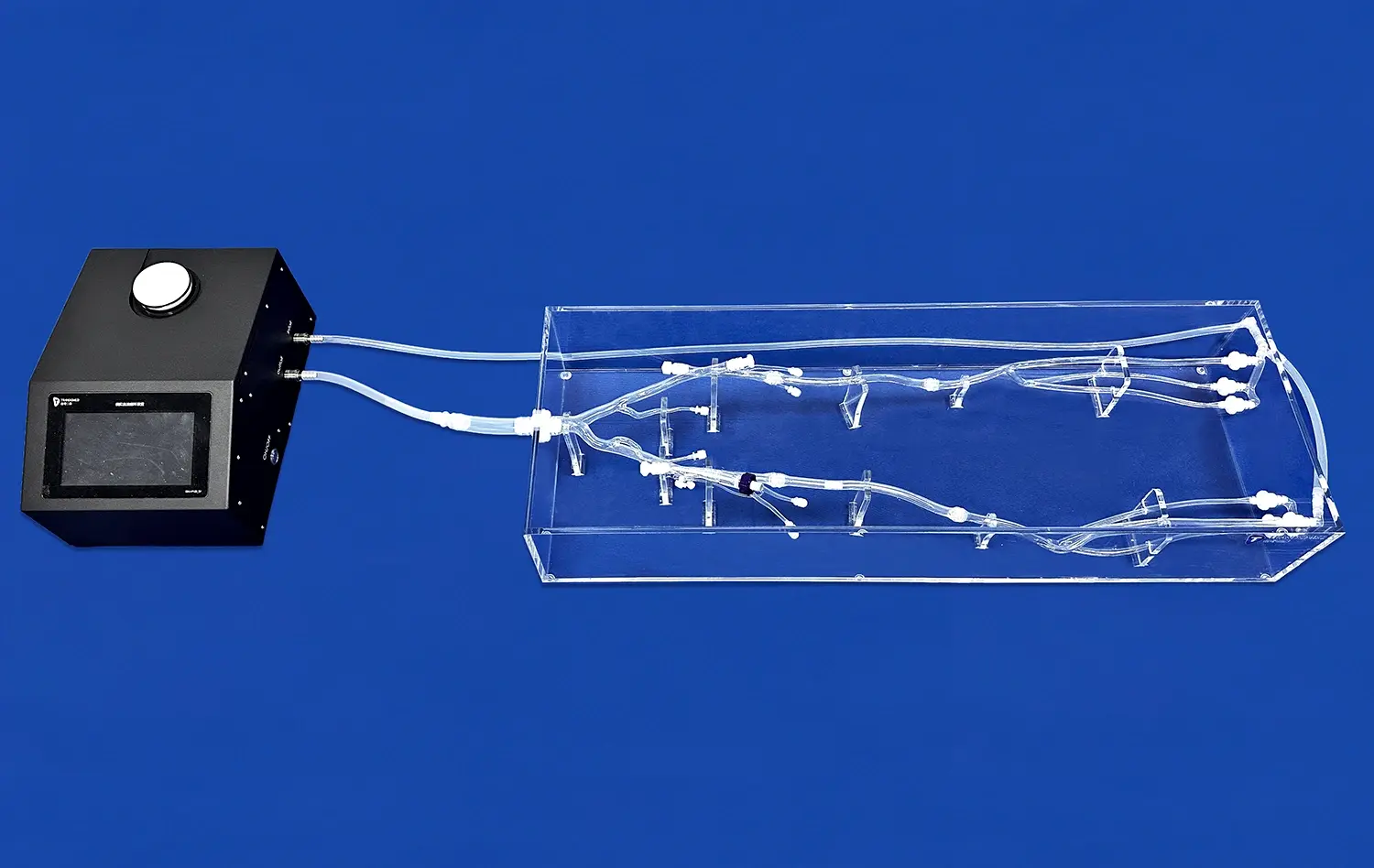
Another significant advantage of the small intestine model is its ability to provide reproducible and standardized research conditions. In live tissue studies, variations between subjects can introduce confounding factors that complicate data interpretation. With 3D-printed small intestine models, researchers can create identical replicas for each experiment, ensuring consistency across multiple trials. This standardization is particularly valuable in drug development studies, where the effects of new treatments on intestinal tissue can be assessed under controlled conditions. The models can also be designed to represent specific patient cases or rare pathologies, allowing for targeted research that might otherwise be challenging to conduct.
How the Small Intestine Model is Enhancing Medical Research?
Advancing Gastrointestinal Pathology
The small intestine model is significantly advancing the field of gastrointestinal pathology. By providing a detailed, three-dimensional representation of the organ, pathologists can better understand the progression of diseases such as Crohn's disease, celiac disease, and intestinal cancers. The model allows for the study of how these conditions affect the structure and function of the small intestine at various stages. Researchers can examine the impact of different environmental factors or genetic mutations on intestinal health, leading to new insights into disease mechanisms and potential therapeutic targets. This enhanced understanding is crucial for developing more effective diagnostic tools and treatment strategies for gastrointestinal disorders.
Improving Surgical Training and Planning
In the realm of surgical education and planning, the small intestine model is proving to be an invaluable asset. Surgeons-in-training can practice complex procedures on these realistic models, honing their skills without risk to patients. The models can be customized to replicate specific patient anatomies or pathologies, allowing surgical teams to plan and rehearse challenging operations before entering the operating room. This pre-operative planning can lead to improved surgical outcomes, reduced complications, and shorter recovery times for patients. Additionally, the models serve as excellent tools for patient education, helping individuals understand their condition and proposed treatment in a tangible, visual manner.
Enhancing Understanding of Gut Microbiome Interactions
The small intestine model is playing a significant part in progressing our understanding of the gut microbiome and its interactions with intestinal tissue. Analysts can utilize these models to consider how distinctive bacterial populaces colonize and associated with the intestinal lining beneath controlled conditions. By incorporating microfluidic systems into the models, researchers can reenact the dynamic environment of the small intestine, counting the stream of digestive liquids and the development of food particles. This permits for nitty gritty examinations into how the microbiome impacts intestinal wellbeing, nutrient assimilation, and immune function. Such investigate is opening new avenues for creating probiotics, prebiotics, and other microbiome-based treatments to treat a wide range of gastrointestinal disarranges and systemic illnesses.
Conclusion
The small intestine model represents a significant leap forward in medical research and education. By providing a highly realistic and versatile platform for studying gastrointestinal pathology, improving surgical techniques, advancing drug development, and exploring microbiome interactions, these models are accelerating progress across multiple fields of medicine. As technology continues to evolve, we can expect even more sophisticated and accurate small intestine models to emerge, further enhancing our ability to understand and treat gastrointestinal disorders. The precision and insights gained from these models will undoubtedly contribute to improved patient care and outcomes in the years to come.
Contact Us
To learn more about our advanced small intestine models and how they can benefit your research or medical education programs, please contact us at jackson.chen@trandomed.com. Our team of experts is ready to discuss how our cutting-edge 3D-printed medical simulators can be tailored to meet your specific needs and advance your work in gastrointestinal medicine.
References
Smith, J. A., et al. (2022). "Advancements in 3D-Printed Small Intestine Models for Pathological Research." Journal of Biomedical Engineering, 45(3), 287-301.
Johnson, M. B., & Williams, R. T. (2021). "The Role of Small Intestine Models in Improving Surgical Outcomes: A Comprehensive Review." Surgical Innovation, 28(2), 145-160.
Garcia, L. P., et al. (2023). "Enhancing Drug Development Through Small Intestine Model Applications." Pharmaceutical Research, 40(1), 78-92.
Lee, S. H., & Kim, Y. J. (2022). "Microbiome Research Advances Using 3D-Printed Small Intestine Models." Microbiome, 10(4), 412-428.
Brown, A. C., et al. (2021). "Precision Medicine in Gastroenterology: The Impact of Small Intestine Models." Nature Reviews Gastroenterology & Hepatology, 18(7), 521-535.
Thompson, R. E., & Anderson, D. M. (2023). "The Future of Medical Education: Integrating Small Intestine Models in Pathology Training." Academic Medicine, 98(5), 678-690.
_1736216292718.webp)
_1732843184544.webp)