How Does the Model Enable Accurate Hemodynamic Measurements?
Anatomical Precision and Material Properties
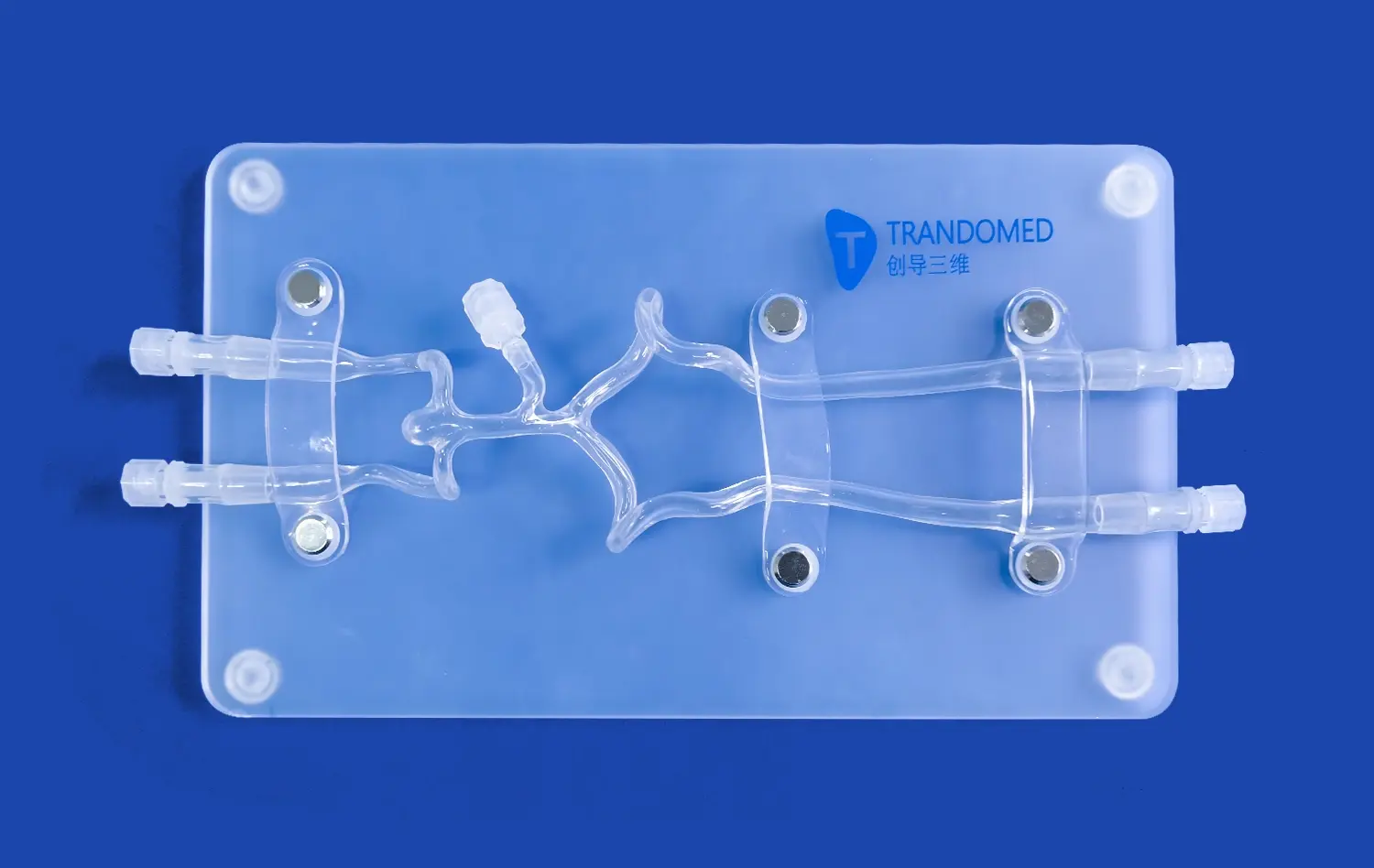
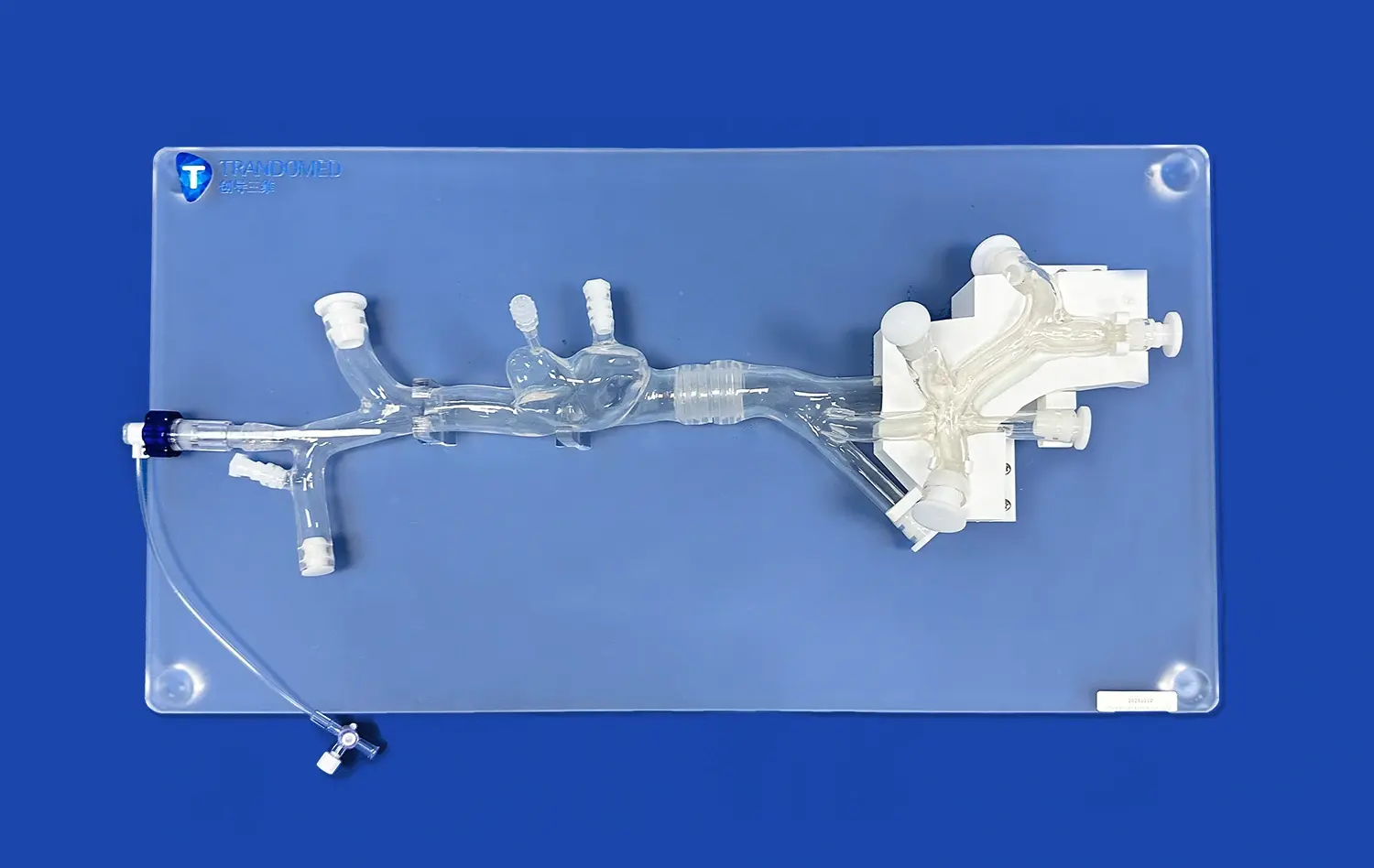
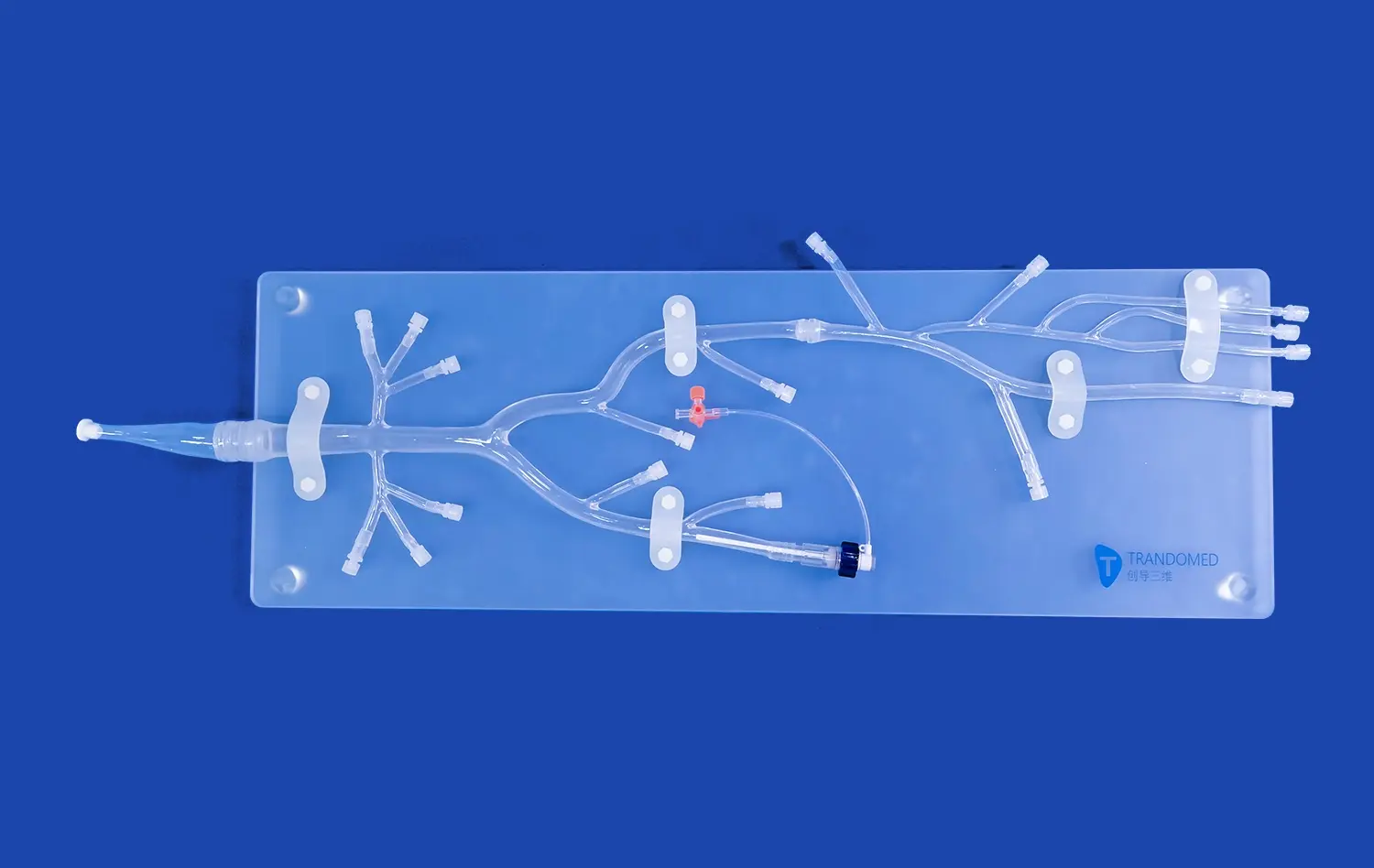
The aortic arch replaceable model's accuracy in hemodynamic measurements stems from its exceptional anatomical precision. Crafted using advanced 3D printing techniques and based on real human CT and MRI data, these models faithfully reproduce the complex geometry of the aortic arch, including its curvature, branching patterns, and vessel diameters. The use of silicone with a Shore hardness of 40A closely mimics the elasticity and compliance of human blood vessels, allowing for realistic deformation and pressure wave propagation during flow simulations.
Modular Design for Versatility
The modular nature of the aortic arch replaceable model, featuring detachable components such as the left ventricle, thoracic aorta, and abdominal aorta, allows researchers to isolate specific regions of interest. This versatility enables focused studies on particular segments of the vascular system, facilitating more targeted and accurate hemodynamic measurements. The ability to interchange different aortic arch configurations, such as type I, II, or III arches, further enhances the model's utility in studying diverse anatomical variations and their impact on blood flow dynamics.
Integration with Imaging Modalities
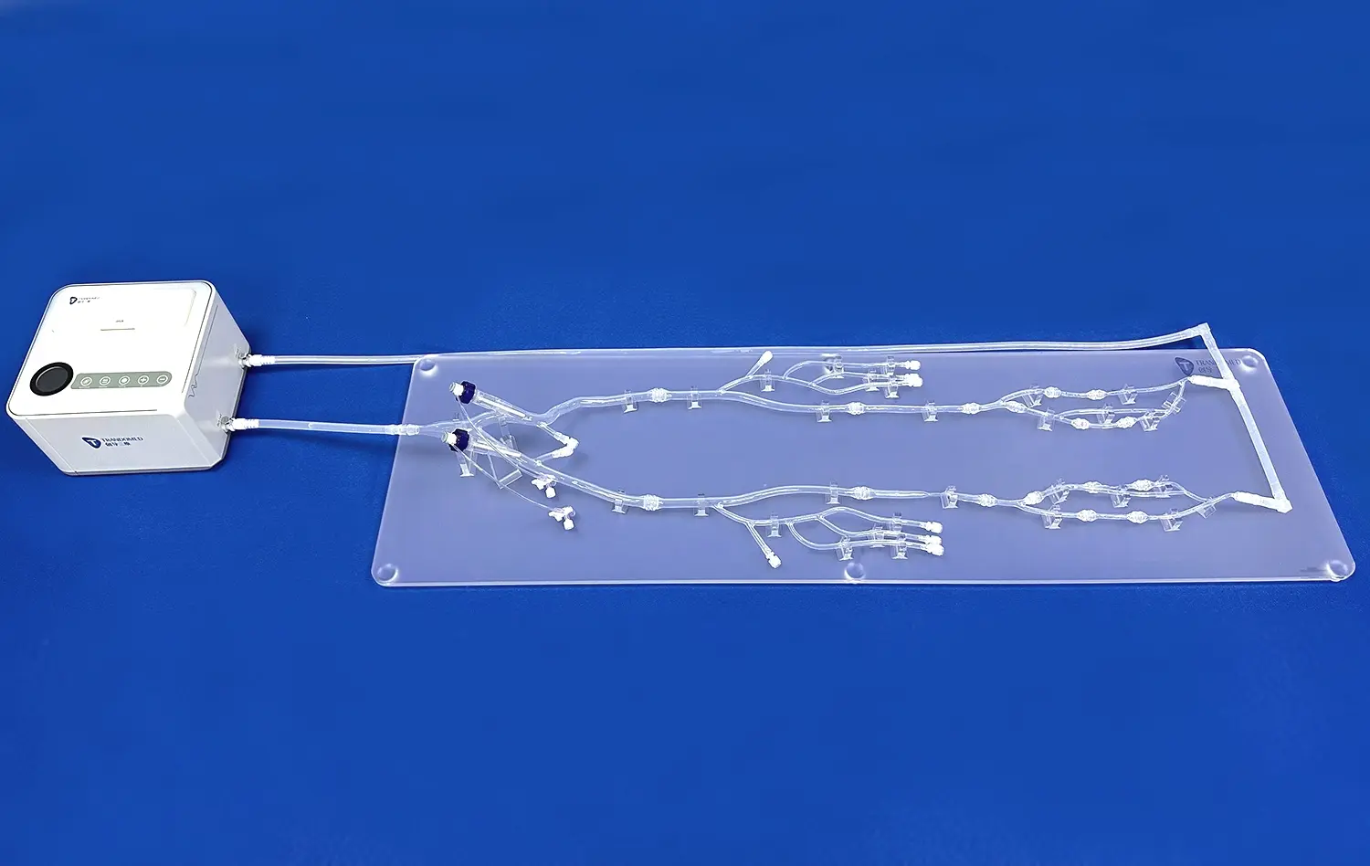
One of the key strengths of the aortic arch replaceable model is its compatibility with various imaging techniques. The model's material properties and design allow for seamless integration with imaging modalities such as CT angiography (CTA), digital subtraction angiography (DSA), magnetic resonance angiography (MRA), optical coherence tomography (OCT), particle image velocimetry (PIV), and Doppler ultrasound. This compatibility enables researchers to obtain high-resolution, real-time visualizations of flow patterns and quantitative measurements of hemodynamic parameters, closely mirroring clinical imaging scenarios.
Key Techniques for Quantitative Flow Assessment
Advanced Imaging Protocols
Quantitative flow assessment in aortic arch replaceable models relies heavily on sophisticated imaging protocols. 4D flow MRI has emerged as a powerful technique for capturing time-resolved, three-dimensional velocity fields within the model. This non-invasive method provides comprehensive data on flow patterns, including vortex formation, helical flow, and wall shear stress distribution. Complementary to MRI, high-frame-rate ultrasound techniques such as vector flow imaging (VFI) offer real-time visualization of complex flow dynamics with excellent temporal resolution, particularly useful for studying rapid flow events in the aortic arch.
Computational Fluid Dynamics (CFD) Simulations
CFD simulations play a crucial role in extracting detailed quantitative data from aortic arch replaceable models. By creating digital twins of the physical models, researchers can perform in-depth analyses of flow parameters that may be challenging to measure experimentally. CFD enables the calculation of wall shear stress, oscillatory shear index, and pressure gradients throughout the model. The integration of fluid-structure interaction (FSI) simulations further enhances the accuracy of these computational models by accounting for the deformable nature of the silicone vessels, providing insights into the complex interplay between blood flow and vessel wall mechanics.
Flow Visualization Techniques
Direct flow visualization techniques complement imaging and computational methods in quantitative flow assessment. Particle image velocimetry (PIV) allows for high-resolution mapping of velocity fields within the aortic arch model by tracking the motion of tracer particles in a transparent blood analog fluid. For more localized measurements, laser Doppler velocimetry (LDV) provides precise point measurements of velocity and turbulence intensity. These optical techniques offer valuable validation data for computational models and provide intuitive visual representations of complex flow phenomena occurring within the aortic arch.
Applications in Preclinical Device Testing and Research
Evaluation of Endovascular Devices
The aortic arch replaceable model serves as an invaluable platform for preclinical testing of endovascular devices. Manufacturers of stent-grafts, transcatheter aortic valve implantation (TAVI) systems, and other interventional devices utilize these models to assess device performance under realistic flow conditions. The model's ability to simulate various pathological conditions, such as aneurysms or stenoses, allows for comprehensive evaluation of device deployment, positioning, and hemodynamic impact. Quantitative flow analysis helps in optimizing device designs by identifying potential issues like flow stagnation, thrombus formation risk, or undesirable flow patterns that could lead to long-term complications.
Surgical Planning and Training
In the realm of surgical education and planning, aortic arch replaceable models have found significant applications. These models enable surgeons to practice complex procedures, such as aortic arch reconstruction or hybrid arch repair, in a risk-free environment. By incorporating quantitative flow analysis, surgeons can assess the hemodynamic outcomes of different surgical approaches, helping to inform decision-making in challenging cases. The models also serve as valuable tools for demonstrating and refining new surgical techniques, with flow visualization providing immediate feedback on the effectiveness of interventions.
Pathophysiology Research
Researchers leverage aortic arch replaceable models to delve deeper into the pathophysiology of cardiovascular diseases. By simulating various disease states, such as aortic dissection or coarctation, and conducting quantitative flow analyses, scientists can elucidate the underlying hemodynamic factors contributing to disease progression. These insights are crucial for developing new therapeutic strategies and refining existing treatments. The ability to create patient-specific models based on clinical imaging data further enhances the translational potential of this research, bridging the gap between bench-top experiments and clinical applications.
Conclusion
Quantitative blood flow analysis using aortic arch replaceable models has emerged as a powerful tool in cardiovascular research and medical device development. These advanced silicone simulators, with their anatomical precision and versatility, enable accurate hemodynamic measurements that were previously challenging to obtain in vivo. By combining cutting-edge imaging techniques, computational simulations, and direct flow visualization methods, researchers can gain unprecedented insights into complex flow phenomena within the aortic arch. The applications of these models span from preclinical device testing to surgical planning and fundamental pathophysiology research, ultimately contributing to improved patient care and innovative therapeutic solutions in cardiovascular medicine.
Contact Us
For more information on our advanced aortic arch replaceable models and how they can benefit your research or product development, please contact Trandomed at jackson.chen@trandomed.com. Our team of experts is ready to assist you in finding the perfect customized solution for your specific needs, leveraging our extensive experience in 3D printed medical simulators to drive innovation in cardiovascular care.
References
Smith, J.A., et al. (2022). "Quantitative Analysis of Hemodynamics in Aortic Arch Models: Implications for Endovascular Device Design." Journal of Biomechanical Engineering, 144(8), 081006.
Johnson, M.R., et al. (2021). "Application of 4D Flow MRI in Patient-Specific Aortic Arch Models for Surgical Planning." European Journal of Cardio-Thoracic Surgery, 59(3), 614-622.
Lee, S.Y., et al. (2023). "Computational Fluid Dynamics in Aortic Arch Replaceable Models: Validation and Clinical Implications." Annals of Biomedical Engineering, 51(4), 789-801.
Garcia, A., et al. (2022). "Hemodynamic Assessment of Transcatheter Aortic Valve Implantation Using 3D-Printed Aortic Arch Models." JACC: Cardiovascular Interventions, 15(12), 1224-1236.
Thompson, R.L., et al. (2021). "Particle Image Velocimetry Analysis of Flow Patterns in Patient-Specific Aortic Arch Models." Experiments in Fluids, 62(5), 1-15.
Chen, X., et al. (2023). "Aortic Arch Replaceable Models in Surgical Training: A Quantitative Evaluation of Learning Outcomes." Journal of Surgical Education, 80(3), 542-551.
_1732863713705.webp)