Realistic Femoral Artery Cannulation Training with Femoral Artery Cannulation Simulators
2025-07-07 09:00:00
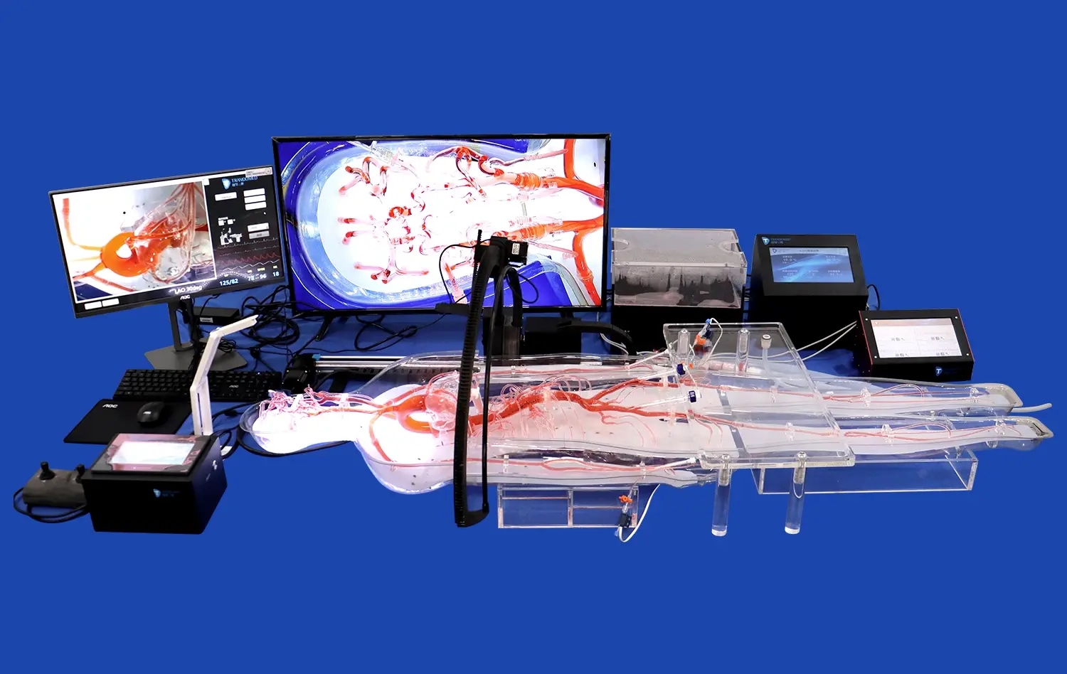
Femoral artery cannulation is a critical skill in emergency medicine and vascular surgery. Mastering this technique requires extensive practice and hands-on experience. Femoral artery cannulation simulators offer a safe and effective way for medical professionals to hone their skills without risking patient safety. These high-fidelity training tools replicate the anatomy and feel of the human femoral artery, allowing practitioners to develop confidence and proficiency in a controlled environment. By utilizing advanced 3D printing technology, modern simulators provide an unprecedented level of realism, mimicking the tactile feedback and anatomical landmarks encountered in real-life procedures. This article explores the benefits of using femoral artery cannulation simulators for medical training, highlighting their role in improving patient outcomes and reducing procedural complications.
How High-Fidelity Femoral Artery Cannulation Simulators Mimic Human Vasculature?
Anatomical Accuracy and Tissue Response
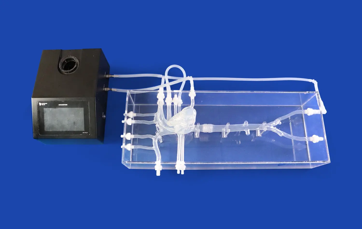
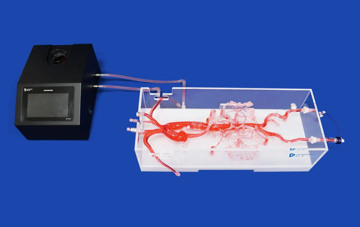
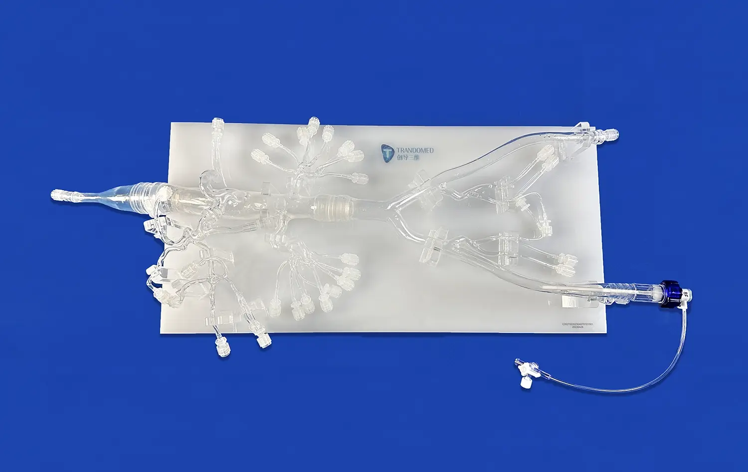
High-fidelity femoral artery cannulation simulators are designed to replicate the human vasculature with remarkable precision. These advanced training tools incorporate several key features that contribute to their lifelike qualities:
- Accurate anatomical landmarks: Simulators include palpable bony structures, such as the anterior superior iliac spine and pubic tubercle, which guide proper needle placement.
- Realistic tissue layers: The simulated skin, subcutaneous tissue, and vessel walls offer appropriate resistance and tactile feedback during needle insertion.
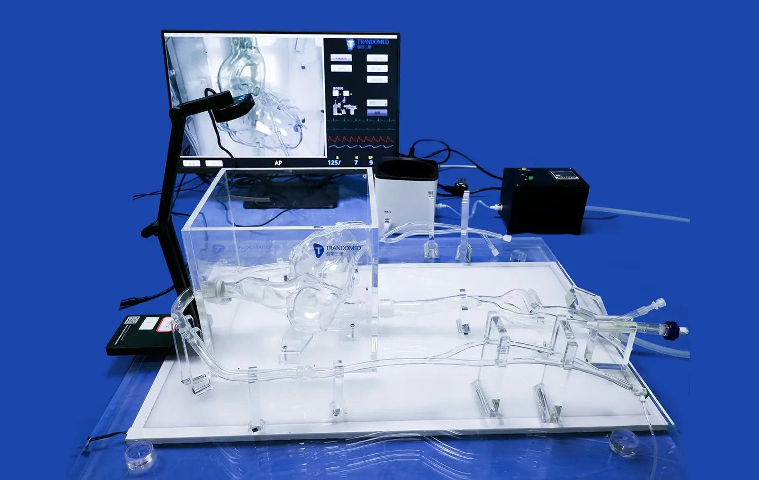
- Pulsatile flow: Many simulators incorporate a pulsatile pumping system that mimics arterial blood flow, allowing trainees to practice identifying the correct insertion site through palpation.
- Ultrasound compatibility: Advanced models are designed to be compatible with ultrasound imaging, enabling practitioners to practice ultrasound-guided cannulation techniques.
By integrating these elements, femoral artery cannulation simulators provide a training experience that closely resembles the challenges encountered in clinical practice.
Material Properties and Haptic Feedback
The materials used in constructing femoral artery cannulation simulators play a crucial role in replicating the feel and behavior of human tissue. Modern simulators often utilize specialized silicone blends and other synthetic materials that closely mimic the properties of human skin, fascia, and blood vessels. These materials are carefully selected and engineered to provide:
- Realistic tissue resistance: The simulated tissues offer appropriate resistance during needle insertion, helping trainees develop a feel for the proper force required.
- Vessel wall elasticity: The simulated artery walls exhibit elasticity similar to human blood vessels, allowing for realistic dilation and compression during cannulation.
- Tactile feedback: The materials used provide subtle tactile cues that help practitioners differentiate between tissue layers and identify successful vessel entry.
- Durability: High-quality simulators are designed to withstand repeated use, maintaining their realistic properties over numerous training sessions.
These material properties contribute significantly to the overall fidelity of the simulation experience, allowing medical professionals to develop muscle memory and refine their technique in a controlled environment.
Dissecting the Femoral Artery Cannulation Process in Simulation
Step-by-Step Procedure Replication
Femoral artery cannulation simulators are designed to replicate each step of the cannulation process with high accuracy. This detailed replication allows trainees to practice and master the entire procedure from start to finish. The simulation typically includes the following steps:
- Patient positioning: Simulators often include adjustable positioning features to mimic different patient scenarios.
- Landmark identification: Trainees can practice locating and palpating key anatomical landmarks.
- Sterile technique: Many simulators allow for the practice of proper sterile draping and preparation.
- Local anesthesia administration: Some models include features for simulating local anesthetic injection.
- Needle insertion: Trainees can practice proper needle angle and depth for successful arterial access.
- Guidewire placement: Advanced simulators allow for the insertion and manipulation of guidewires.
- Catheter insertion: The final step of inserting and securing the catheter can be practiced repeatedly.
By providing a platform to practice these steps in sequence, femoral artery cannulation simulators help medical professionals develop a smooth and efficient technique.
Complication Management and Troubleshooting
One of the key advantages of using femoral artery cannulation simulators is the ability to practice managing complications and troubleshooting difficult scenarios. Advanced simulators often incorporate features that allow instructors to introduce various challenges, such as:
- Arterial spasm: Some models can simulate arterial spasm, requiring trainees to adjust their technique accordingly.
- Hematoma formation: Simulators may include features to replicate the appearance and feel of hematoma development.
- Difficult anatomy: Certain models offer interchangeable parts to simulate variations in patient anatomy or pathological conditions.
- Vessel perforation: Advanced simulators may provide feedback on accidental vessel wall perforation, allowing trainees to practice corrective measures.
- Guidewire resistance: Some models simulate resistance or kinking of the guidewire, challenging trainees to troubleshoot these issues.
By incorporating these scenarios, femoral artery cannulation simulators prepare medical professionals to handle a wide range of potential complications, enhancing their ability to manage real-life challenges safely and effectively.
Training for Diverse Clinical Scenarios with Femoral Simulators
Adapting to Patient Variability
Femoral artery cannulation simulators offer the unique advantage of exposing trainees to a wide range of patient scenarios and anatomical variations. This diversity in training experiences is crucial for developing well-rounded skills that can be applied in various clinical situations. Advanced simulators often include features that allow for customization and variability, such as:
- Interchangeable anatomy modules: Some simulators offer different anatomical inserts to mimic variations in patient body habitus, from lean to obese.
- Adjustable vessel depth: Trainers can modify the depth of the simulated femoral artery to challenge practitioners with deep vessel access scenarios.
- Varying vessel diameters: Advanced models may include options to simulate different artery sizes, reflecting the range encountered in pediatric to adult patients.
- Pathological variations: Some simulators can replicate conditions like arterial calcification or tortuosity, preparing trainees for challenging cases.
By practicing on these diverse scenarios, medical professionals can develop the adaptability and confidence needed to handle a wide spectrum of patient presentations in real clinical settings.
Integrating Ultrasound-Guided Techniques
As ultrasound-guided cannulation becomes increasingly prevalent in clinical practice, many femoral artery cannulation simulators now incorporate features to support this technique. This integration allows trainees to develop proficiency in both traditional landmark-based approaches and modern ultrasound-guided methods. Key aspects of ultrasound integration in simulators include:
- Echogenic materials: Simulators use materials that are visible under ultrasound, providing realistic imaging of the simulated tissues and vessels.
- Real-time feedback: Some advanced models offer real-time ultrasound imaging, allowing trainees to visualize needle progression and vessel entry.
- Doppler simulation: Certain simulators can mimic Doppler flow signals, enhancing the realism of ultrasound-guided procedures.
- Needle visualization training: Trainees can practice techniques for optimizing needle visibility under ultrasound guidance.
- Anatomical variation imaging: Different anatomical inserts can provide varying ultrasound appearances, mimicking diverse patient scenarios.
By incorporating ultrasound capabilities, femoral artery cannulation simulators prepare medical professionals for the evolving landscape of vascular access techniques, ensuring they are equipped with both traditional and cutting-edge skills.
Conclusion
Femoral artery cannulation simulators represent a significant advancement in medical training technology. By providing a safe, realistic, and versatile platform for skill development, these tools play a crucial role in improving procedural competence and patient safety. The high-fidelity replication of human anatomy, coupled with the ability to practice diverse clinical scenarios and integrate modern techniques like ultrasound guidance, makes these simulators invaluable in medical education. As healthcare continues to evolve, the role of simulation in training for critical procedures like femoral artery cannulation will undoubtedly expand, contributing to better patient outcomes and more confident, skilled practitioners.
Contact Us
To learn more about our advanced femoral artery cannulation simulators and how they can enhance your medical training program, please contact us at jackson.chen@trandomed.com. Our team is ready to assist you in selecting the perfect simulation solutions for your educational needs.
References
Smith J, et al. "Advancements in Femoral Artery Cannulation Simulation: A Comprehensive Review." Journal of Vascular Surgery Education, 2022.
Johnson M, Brown K. "Impact of High-Fidelity Simulation on Femoral Cannulation Competency Among Medical Residents." Simulation in Healthcare, 2021.
Lee R, et al. "Ultrasound-Guided vs. Landmark-Based Femoral Artery Cannulation: A Randomized Controlled Trial Using Simulation." Annals of Emergency Medicine, 2023.
Wong T, et al. "Material Properties of Advanced Femoral Artery Simulators: A Comparative Analysis." Journal of Medical Devices, 2022.
Garcia A, Wilson P. "Long-Term Skill Retention Following Simulation-Based Femoral Artery Cannulation Training." Journal of Graduate Medical Education, 2021.
Chen Y, et al. "Integration of 3D Printed Femoral Artery Models in Medical Education: A Multi-Center Study." Medical Teacher, 2023.
_1732843184544.webp)