Realistic Venous Cardiac Anatomy for Electrophysiology Education
2026-05-14 09:00:04
More than just textbooks and diagrams are needed to improve teaching in cardiac electrophysiology. Medical schools, hospitals, and gadget makers all over the world can't do their work without anatomically accurate simulation tools that can copy the heart's complex vein structures. A good venous cardiac electrophysiology model helps students learn both theory and practical skills. It lets them practice ablation techniques, understand how electrical signals travel through the heart, and handle complicated catheter procedures without any risk. As healthcare facilities look for effective training options, it's important to know what these specialized anatomical simulators can do and how they can be used so that institutions can make smart purchasing choices that improve patient safety and education.
Understanding Venous Cardiac Electrophysiology Models
Defining Core Components and Anatomical Accuracy
Venous cardiac electrophysiology models are specialized teaching aids that were made to accurately mimic the heart's venous system. The superior vena cava, inferior vena cava, right atrium, right ventricle, and subclavian vein are just a few of the important anatomical features that these simulators carefully copy. Unlike whole-heart models, which show the heart as a whole, venous-specific designs focus on the pathways and structures that are important for electrophysiological treatments. This lets professionals get better at specific skills like catheter navigation and mapping.
Distinguishing Venous from Arterial Models
Because venous and arterial heart parts are built differently, they need different modeling methods. Venous models focus on capacitance vessels with thin walls and lower pressure, while arterial models have thicker walls and pulsatile flow patterns. In EP studies, this difference has a big effect on how realistic the simulations are because catheter behavior, guidewire advancement, and device deployment are all very different between venous and arterial access paths. Medical training programs can learn a lot from venous simulators that correctly replicate the tactile feedback and anatomical landmarks that doctors see during real treatments.
Applications Across Medical Training and Research
Realistic venous anatomy models are useful for many people in the healthcare system. These models are used by medical schools to teach basic heart anatomy and get students comfortable with interventional methods before they work with real patients. High-fidelity simulators are used by hospital training departments to certify staff in advanced EP procedures. This lowers the number of complications and increases the efficiency of the operations. Anatomically accurate models are used by medical device companies to test their products, send them to regulators, and show them to potential customers. Research labs also use them for biomechanical studies and to confirm new techniques.
Comparison of Venous Cardiac Electrophysiology Models and Solutions
High-Fidelity Versus Simplified Educational Models
There is a wide range of venous electrophysiology simulations on the market, from simple models of the body to complex, high-fidelity systems. Simplified models give you a basic idea of how bodies work and are good for learning basic skills in schools that don't have a lot of money. On the other hand, high-fidelity solutions use materials that are similar to tissue, have true tactile properties, and include complex anatomical variations that reflect clinical diversity. When making choices about what simulators to buy, the level of difficulty should match the training goals. For example, simplified versions may work well for introductory anatomy classes, but surgical skills labs and device testing facilities need more advanced models that accurately replicate clinical conditions.
Physical Models Versus Computational Simulations
There are two different but related ways to teach EP: physical models made of silicone and computer simulations. Physical models give learners invaluable tactile feedback, which helps them develop the skills they need to manipulate catheters. The three-dimensional spatial knowledge that comes from touching and interacting with real body parts can't be fully matched by learning on a screen. Computational models are great at showing how electricity flows and modeling rare diseases, but they lack the tactile aspect that is necessary for learning how to do things. More and more, comprehensive training programs use both of these methods together to make learning more effective.
Material Considerations and Durability
The materials used to build venous heart models have a big effect on how useful they are for teaching and how long they last. In high-end models, Silicone Shore 40A is often used because it nearly matches the properties of human tissue and is very durable for repeated catheter insertions. This material keeps its properties the same after thousands of training sessions, giving reliable results that justify business investment. Alternative materials may be cheaper to buy at first, but they often need to be replaced too soon because they break down, which raises the total cost of ownership for training sites that run a lot of educational programs.
Development and Simulation Techniques for Venous Cardiac Electrophysiology Models
Data Acquisition and Anatomical Reconstruction
Getting a lot of medical imaging data is the first step in making physically accurate venous cardiac models. For correct anatomical reconstruction, high-resolution CT and MRI scans from a wide range of patients are needed. This imaging data is processed by reverse 3D reconstruction technology to get a thorough picture of the venous architecture. This includes differences in the diameter of each vessel, its branching patterns, and its spatial relationships. This method, which is based on data, makes sure that training venous cardiac electrophysiology models show real human anatomy instead of artificial textbook versions. This helps students get ready for the wide range of anatomy they will see in clinical practice.
Advanced Manufacturing Through 3D Printing
The way cardiac simulation tools are made has changed a lot thanks to new manufacturing methods. Unique 3D printing techniques make it possible to make complex internal shapes and thin-walled venous structures that aren't possible with standard molding methods. With layer-by-layer additive manufacturing, you can precisely control the model's wall thickness, lumen width, and tissue compliance. This level of accuracy in manufacturing directly translates to realism in simulations, as catheters meet resistance that is appropriate for the body and navigate accurately reproduced anatomical landmarks during training procedures.
Customization Capabilities for Specialized Training
An important step forward in medical simulation is being able to change anatomical models to meet specific teaching goals. Training programs can make changes based on CT data files in STL, STEP, and CAD formats. This lets them make anatomical models of particular patients for planning procedures before they happen or teaching rare pathology. Adjustable features like foramen ovale sizes, atrial septal variations, and venous anomalies can be added without any design fees. This lets institutions create custom training situations that meet their specific course needs and procedure focus areas.
Procuring and Integrating Venous Cardiac Electrophysiology Modeling Tools
Evaluating Supplier Capabilities and Support
To find a reliable supplier for cardiac simulation equipment, you need to carefully consider more than just the product specs. Supplier experience in making medical devices shows how mature the process is and how consistent the quality is. Technical support skills show how well institutions can solve practical problems. Manufacturers that have been specializing in medical 3D printing technology for decades bring a lot of useful knowledge to the table when it comes to developing new products and can help you choose the best models for your training needs. Customer service that responds quickly, short wait times, and detailed documentation make it easy to add simulation tools to training programs that are already in place.
Cost Analysis and Return on Investment
When you buy good venous electrophysiology models, you get measured returns in a number of ways. Training programs lower the number of complications by giving doctors and nurses time to learn methods before they do procedures on patients. This directly improves patient safety and lowers the risk of malpractice. Medical device companies shorten the time it takes to make a new product by trying early versions on anatomical models instead of expensive animal studies or human trials that are delayed. The long life of high-quality silicone models spreads the cost of purchase over thousands of training sessions, resulting in much lower per-procedure costs compared to alternatives that are used up quickly or cadaveric examples that are hard to get and raise ethical concerns.
Logistics and Operational Integration
When putting simulation tools to use, practical procurement issues play a role. Institutional buying processes and spending cycles can be accommodated by flexible payment terms, such as T/T transactions. Fast lead times of seven to ten days make it possible to start the program quickly, and FedEx, DHL, EMS, UPS, and TNT all offer reliable international shipping choices to training facilities all over the world. Because physical models are small and self-contained, they don't need to be installed in a complicated way. This means that they can be used right away after delivery without the need for special infrastructure or expert help during setup.
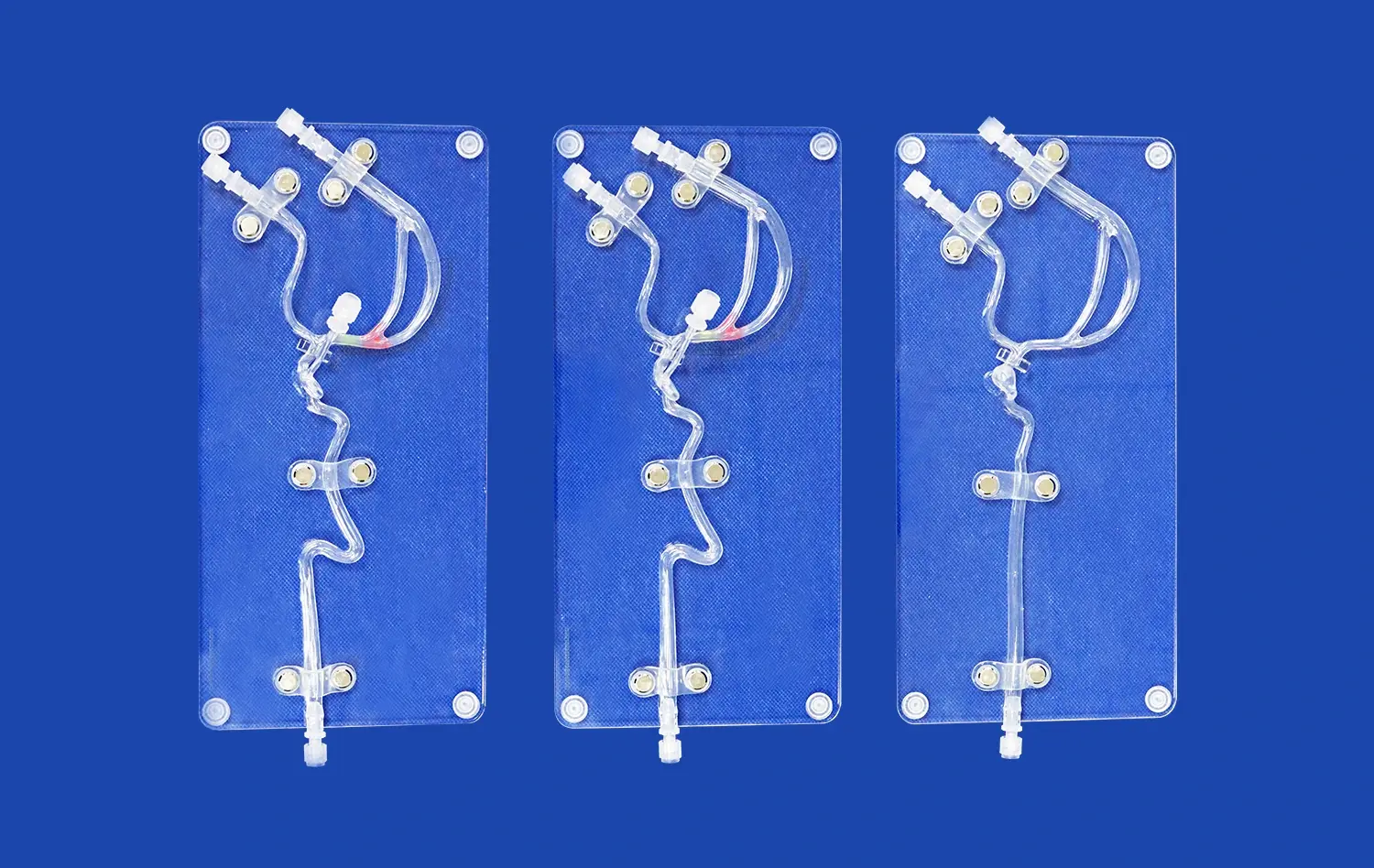
The Trandomed XXS004 Venous Cardiac Electrophysiology Model
For training to be excellent, simulation tools must accurately reflect actual situations. The Venous Cardiac Electrophysiology Model (Product No. XXS004) is an example of how precision engineering can be used to teach medicine. It is made from Silicone Shore 40A and has anatomical structures that are carefully created. Based on large CT and MRI files of real people, this amazing simulator accurately recreates the inferior vena cava, superior vena cava, right atrium, right ventricle, and subclavian vein.
The design of the model specifically meets the training needs of EP procedures by giving realistic experiences of navigating a catheter through venous access routes that are widely used in clinical practice. This simulator has anatomically exact landmarks and tissue-equivalent tactile inputs that help with electrophysiological mapping exercises, developing ablation techniques, and training for deploying devices. Schools have found that students learn how to do things faster when they move from this accurate venous cardiac electrophysiology model to supervised clinical cases. This is because the students' anatomical knowledge and manual skills are easily applied to the patient care setting.
This answer is different from generic ones because it can be customized. Atrial geometry can be rebuilt using institutional CT data. This lets doctors plan pre-procedural steps for each patient or make training scenarios with different body parts. The size of the foramen ovale can be changed to fit different training goals, and the layout of the venous pathway can be changed to fit different ways of doing things. These changes are made without adding extra costs to the design, showing that the company wants to encourage new ideas in education instead of setting strict standards.
The performance traits that training programs count on are based on excellent manufacturing. Unique 3D printing molding methods make sure that delicate anatomical features are always reproduced accurately across production runs, and the material formulation finds the best balance between durability and tissue-equivalent compliance. The model can be used over and over again to insert catheters without breaking down. It keeps its anatomical accuracy through thousands of training procedures and gives reliable results that teachers can safely use in standardized competency tests.
Future Trends and Innovations in Venous Cardiac Electrophysiology Modeling
Integration of Multi-Scale Simulation Approaches
Emerging educational paradigms combine physical anatomical models with computational electrophysiology simulations to create comprehensive learning experiences. Students manipulate catheters within tangible venous structures while simultaneously observing real-time electrical activation patterns displayed on synchronized screens. This multi-modal integration reinforces the relationship between anatomical navigation and electrophysiological phenomena, deepening conceptual understanding while developing procedural skills. As technological capabilities advance, we anticipate increasingly seamless integration between physical simulators and digital visualization systems.
Augmented and Virtual Reality Applications
Immersive technologies promise to enhance traditional simulation methodologies by overlaying additional information onto physical training experiences. Augmented reality systems can project catheter position, electrical activation maps, or anatomical annotations directly onto physical models, enriching the educational context without sacrificing the tactile dimension essential for skill development. Virtual reality applications enable remote collaborative training, allowing expert instructors to guide geographically distributed learners through complex procedures using shared digital representations of standardized anatomical models.
Patient-Specific Modeling for Precision Medicine
The trajectory toward personalized healthcare extends into procedural training and planning. Advanced medical centers increasingly create patient-specific cardiac models from pre-procedural imaging, allowing surgical teams to rehearse complex cases using exact anatomical replicas before the actual intervention. This approach reduces procedural time, minimizes complications, and improves outcomes for patients with challenging anatomy. As manufacturing costs decline and turnaround times compress, patient-specific modeling will transition from exceptional cases to routine practice, fundamentally transforming pre-procedural preparation protocols.
Strategic Recommendations for Adoption
Organizations seeking to maintain competitive advantages in medical education and device development should proactively invest in advanced simulation capabilities. Establishing relationships with experienced manufacturers ensures access to emerging technologies and customization expertise as training requirements evolve. Institutional commitment to simulation-based education, supported by appropriate equipment procurement and faculty development, generates measurable improvements in learner competency and patient safety metrics that justify continued investment in these transformative educational tools.
Conclusion
Realistic venous cardiac anatomy simulators have become foundational elements of modern electrophysiology education, enabling safe skill development and accelerating practitioner competency across diverse healthcare settings. The integration of data-driven anatomical reconstruction, advanced manufacturing techniques, and customization capabilities creates simulation tools that faithfully replicate clinical realities. As medical training increasingly emphasizes hands-on procedural competence, procurement decisions regarding anatomical venous cardiac electrophysiology models directly impact educational effectiveness and ultimately patient care quality. Organizations that strategically invest in high-fidelity simulation equipment position themselves at the forefront of medical education innovation, preparing the next generation of electrophysiologists with the knowledge and skills necessary for clinical excellence.
FAQ
What distinguishes venous from arterial cardiac electrophysiology models?
Venous models specifically replicate the thin-walled, low-pressure vessels used for catheter access during EP procedures, including the superior and inferior vena cavae, subclavian veins, and right heart chambers. Arterial models feature thicker walls and different mechanical properties. Training realism improves when simulators accurately represent the specific vascular territories relevant to particular procedures.
How can institutions ensure simulation accuracy when selecting models?
Verification that models derive from actual human CT and MRI data rather than idealized drawings ensures anatomical authenticity. Material properties should approximate human tissue compliance, and manufacturers with extensive experience in medical 3D printing typically deliver superior accuracy. Requesting customization based on institutional imaging data provides the highest fidelity for specialized training scenarios.
What factors influence the total cost of ownership for cardiac simulation equipment?
Beyond initial acquisition expense, durability determines replacement frequency and long-term costs. Premium silicone models withstand thousands of training sessions, while inferior materials degrade rapidly. Supplier support, customization availability without design fees, and rapid lead times also contribute value. Comprehensive cost analysis should evaluate per-procedure expenses across the model's functional lifespan rather than focusing exclusively on purchase amounts.
Partner with a Trusted Venous Cardiac Electrophysiology Model Manufacturer
Trandomed specializes in developing anatomically precise simulation tools that elevate medical education standards worldwide. Our Venous Cardiac Electrophysiology Model (XXS004) combines two decades of medical 3D printing expertise with manufacturing excellence, delivering training solutions that healthcare institutions, device manufacturers, and research laboratories depend upon for procedural education and product development. Customization capabilities, expedited production timelines, and comprehensive technical support ensure that your specific requirements receive expert attention from initial consultation through deployment. Connect with our team at jackson.chen@trandomed.com to discuss how our venous cardiac electrophysiology model supplier capabilities can advance your training programs, device testing protocols, or research initiatives with precision simulation tools engineered for your success.
References
Josephson, M.E. (2016). Clinical Cardiac Electrophysiology: Techniques and Interpretations. Philadelphia: Wolters Kluwer Health.
Zipes, D.P., Jalife, J., & Stevenson, W.G. (2018). Cardiac Electrophysiology: From Cell to Bedside. Philadelphia: Elsevier Saunders.
Haïssaguerre, M., & Shah, D.C. (2019). Venous Access and Navigation Techniques in Complex Electrophysiology Procedures. Journal of Interventional Cardiac Electrophysiology, 54(2), 127-138.
Moorman, A.F., & Christoffels, V.M. (2017). Cardiac Chamber Formation: Development, Genes, and Evolution. Physiological Reviews, 97(4), 1297-1336.
Panescu, D. (2020). Medical Simulation Training Systems for Cardiac Electrophysiology Education. IEEE Engineering in Medicine and Biology Magazine, 39(3), 45-52.
Winkle, R.A., Mead, R.H., & Engel, G. (2015). The Role of Anatomical Models in Electrophysiology Training and Procedural Planning. Heart Rhythm Education, 12(6), 892-901.
_1736215128474.webp)
_1734507205192.webp)