What Technologies Are Shaping Next-Generation Heart Models?
Advanced Imaging Techniques
The foundation of accurate heart models lies in high-resolution imaging. Cutting-edge imaging technologies such as 4D cardiac CT and 7T MRI are revolutionizing our ability to capture intricate details of cardiac structures. These modalities provide unprecedented spatial and temporal resolution, allowing for the creation of heart models that faithfully represent individual patient anatomies. The integration of multiple imaging techniques, including echocardiography and nuclear imaging, further enhances the comprehensive nature of these models, capturing both structural and functional aspects of the heart.
Artificial Intelligence and Machine Learning
Artificial intelligence (AI) and machine learning algorithms are playing an increasingly crucial role in the development of advanced heart models. These technologies excel at processing vast amounts of imaging data, automatically segmenting cardiac structures, and generating detailed 3D reconstructions. AI-powered models can predict cardiac behavior under various conditions, simulating the effects of different interventions or disease progressions. This predictive capability is particularly valuable for personalized treatment planning and risk assessment.
Biomechanical Modeling
Sophisticated biomechanical modeling techniques are enhancing the realism of heart simulations. These models incorporate complex mathematical equations that describe cardiac tissue properties, fluid dynamics, and electrophysiology. By simulating the intricate interplay between these factors, researchers can create virtual heart models that behave remarkably like their biological counterparts. Such models are invaluable for studying cardiac mechanics, predicting surgical outcomes, and developing new therapeutic approaches for conditions like heart failure or arrhythmias.
3D Printing, Silicone Replication, and Functional Flow Simulation
Advancements in 3D Printing Technology
The realm of 3D printing has opened up new possibilities in creating tangible, highly accurate heart models. Multi-material 3D printing allows for the fabrication of models that mimic the varying tissue properties found in the heart, from the flexible myocardium to the more rigid valvular structures. These models can be customized to replicate specific patient anatomies, providing surgeons with the opportunity to rehearse complex procedures before entering the operating room. The ability to print transparent models also allows for enhanced visualization of internal structures, facilitating a deeper understanding of spatial relationships within the heart.
Silicone-Based Cardiac Replicas
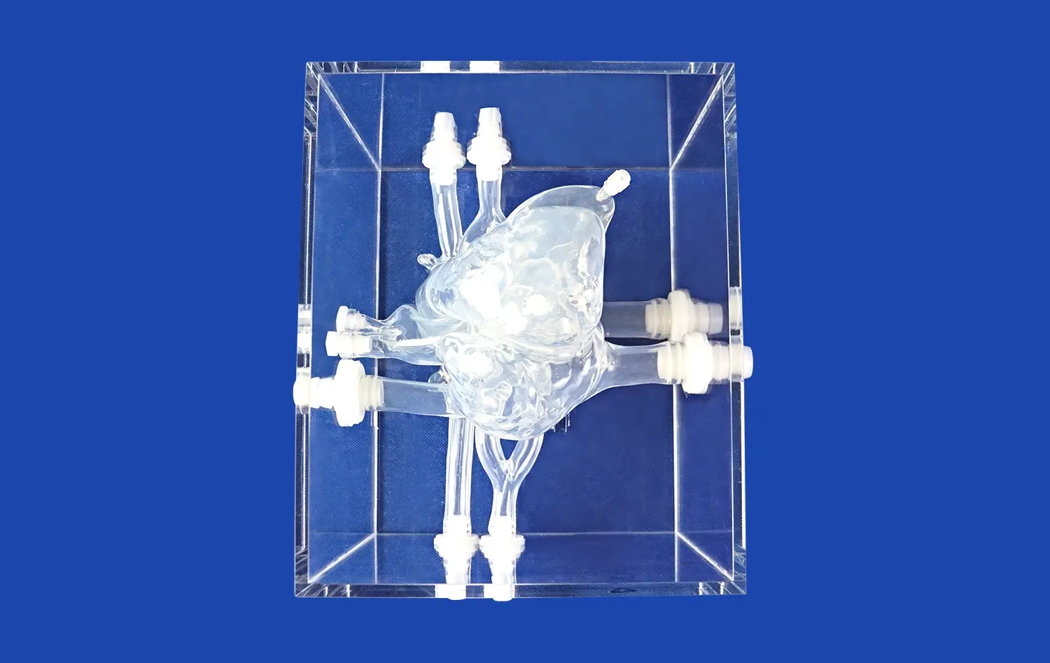
Silicone has emerged as a preferred material for creating lifelike heart models due to its versatility and similarity to human tissue. Companies like Trandomed are at the forefront of this technology, utilizing proprietary 3D printing molding techniques to produce high-fidelity silicone heart models. These models, such as the Heart Model with Coronary (XXK002DJ), offer exceptional detail, including accurate representations of the coronary arteries and major cardiac chambers. The use of silicone Shore 40A material closely mimics the texture and elasticity of cardiac tissue, providing a realistic tactile experience for training and simulation purposes.
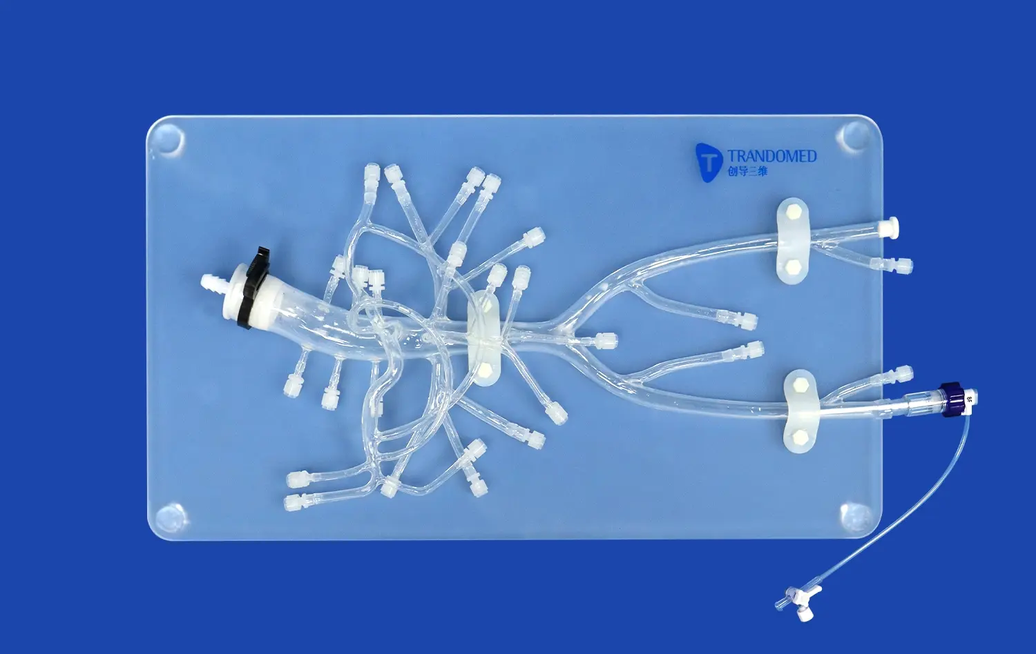
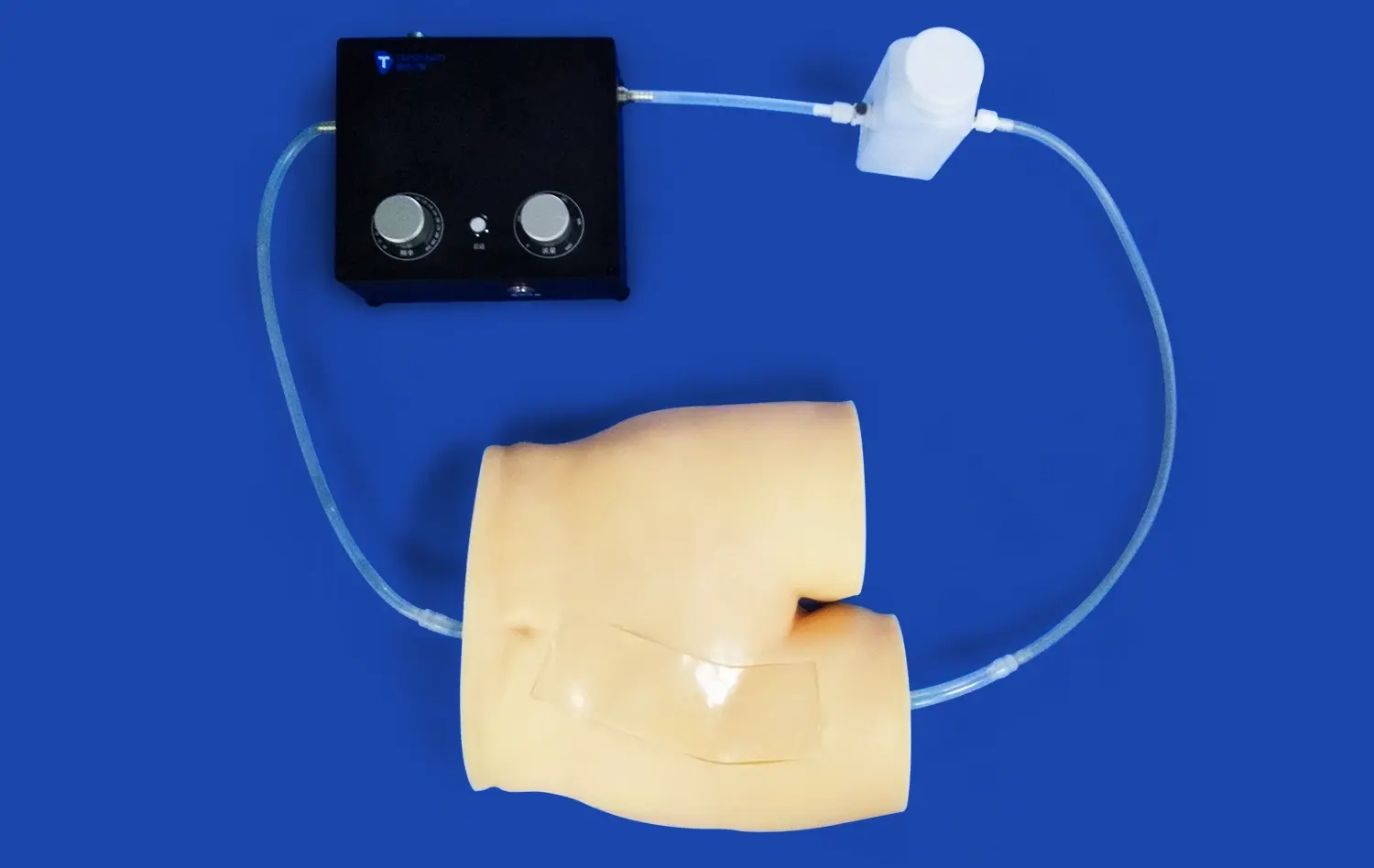
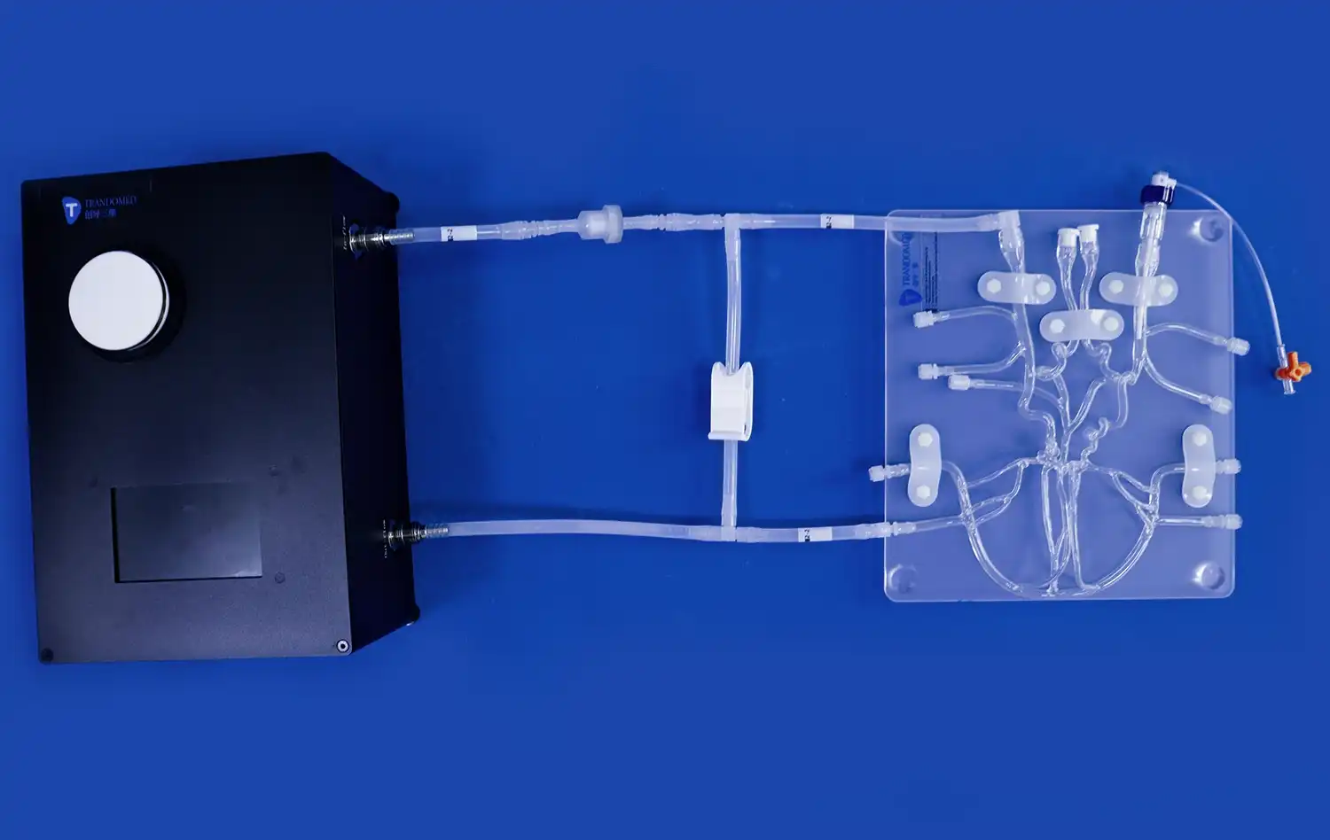
Dynamic Flow Simulation
The integration of functional flow simulation into physical heart models represents a significant leap forward in cardiac simulation technology. These advanced models incorporate channels and reservoirs that allow for the circulation of fluid, mimicking blood flow through the heart and great vessels. This dynamic element enables the simulation of various physiological and pathological conditions, such as valve regurgitation or stenosis. Researchers and clinicians can observe and measure flow patterns, pressure gradients, and other hemodynamic parameters, providing invaluable insights into cardiac function and the effects of interventional procedures.
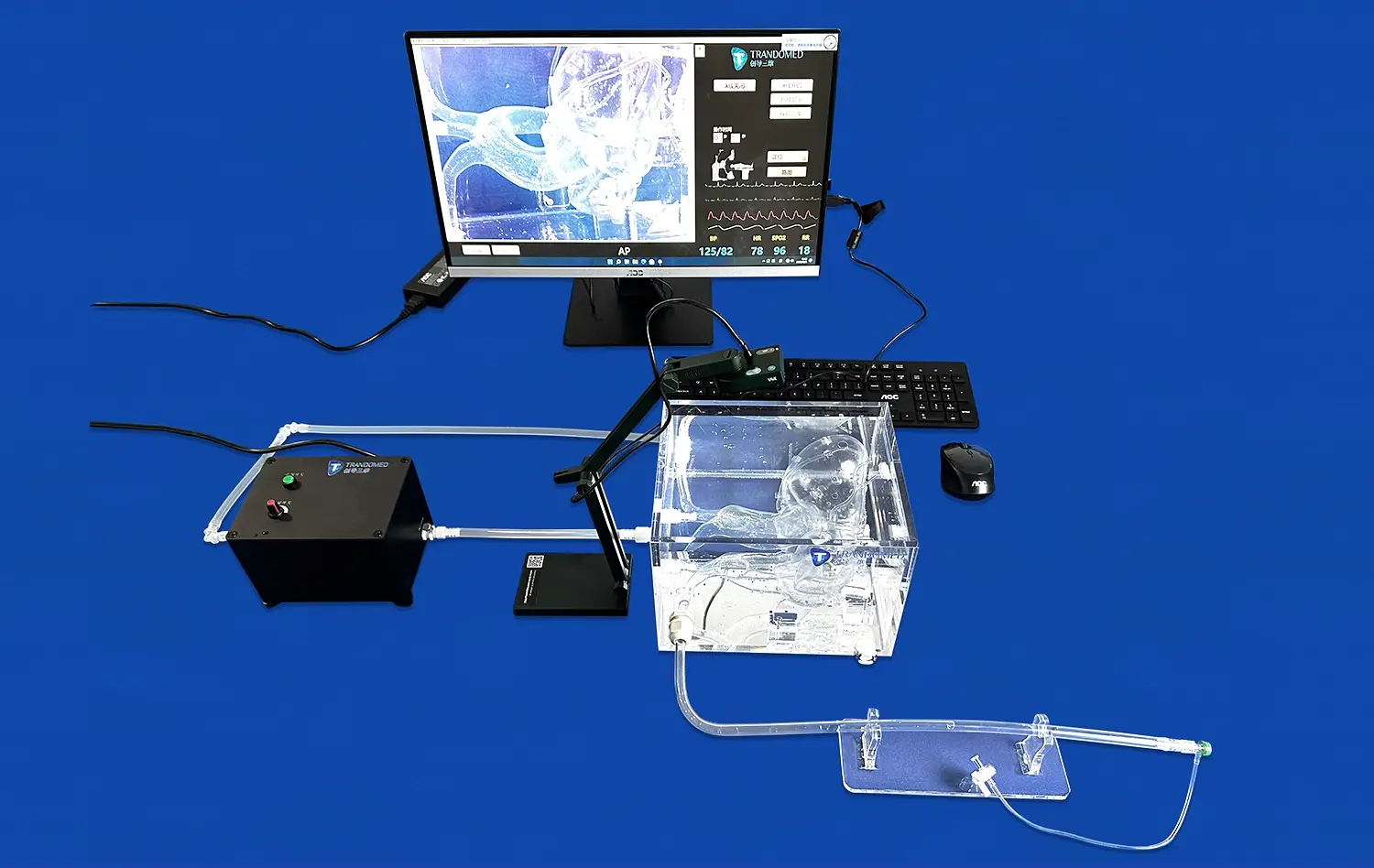
Integration with Imaging and Augmented Reality Systems
Image Fusion and Virtual Reality
The convergence of physical heart models with digital imaging technologies is creating immersive learning environments. Advanced image fusion techniques allow for the seamless integration of patient-specific imaging data with physical models, creating hybrid systems that combine the benefits of tangible interaction with the flexibility of digital manipulation. Virtual reality (VR) platforms take this concept further, enabling users to explore highly detailed, interactive 3D renderings of the heart. These VR simulations can be programmed to respond to user input, simulating the effects of various interventions in real-time.
Augmented Reality in Cardiac Education and Planning
Augmented reality (AR) is transforming how medical professionals interact with heart models. AR systems can overlay digital information onto physical models or project virtual heart models into the real world. This technology allows for dynamic, interactive exploration of cardiac anatomy and function. In educational settings, AR-enhanced heart models can provide students with a more engaging and intuitive learning experience. For surgical planning, AR can be used to visualize potential approaches, identify anatomical landmarks, and simulate procedural outcomes, all within the context of the patient's actual anatomy.
Real-Time Data Integration
The latest advancements in heart modeling technology include the ability to integrate real-time patient data into simulations. This capability allows for the creation of "digital twins" – virtual representations of a patient's heart that can be updated in real-time based on ongoing monitoring and diagnostic information. These dynamic models have the potential to revolutionize patient care by providing continuous, personalized risk assessment and treatment optimization. In the operating room, real-time integration of imaging and physiological data with AR-enhanced heart models could provide surgeons with unprecedented guidance during complex procedures.
Conclusion
The future of cardiac simulation is marked by remarkable advancements in heart modeling technology. From high-fidelity 3D-printed replicas to dynamic digital simulations, these innovations are transforming medical education, research, and patient care. As we continue to push the boundaries of what's possible in cardiac modeling, we can anticipate even more sophisticated tools that will enhance our understanding of heart function and improve outcomes for patients with cardiovascular diseases. The integration of AI, AR, and real-time data analysis promises to usher in a new era of personalized cardiac care, where highly accurate, patient-specific heart models guide every aspect of diagnosis, treatment planning, and intervention.
Contact Us
At Trandomed, we're committed to advancing the field of cardiac simulation through our innovative 3D-printed medical models and simulators. Our expertise in creating highly realistic, customizable heart models positions us at the forefront of this exciting technological revolution. To learn more about how our advanced heart models can enhance your medical training, research, or clinical practice, please contact us at jackson.chen@trandomed.com. Together, we can shape the future of cardiovascular medicine.
_1732843184544.webp)