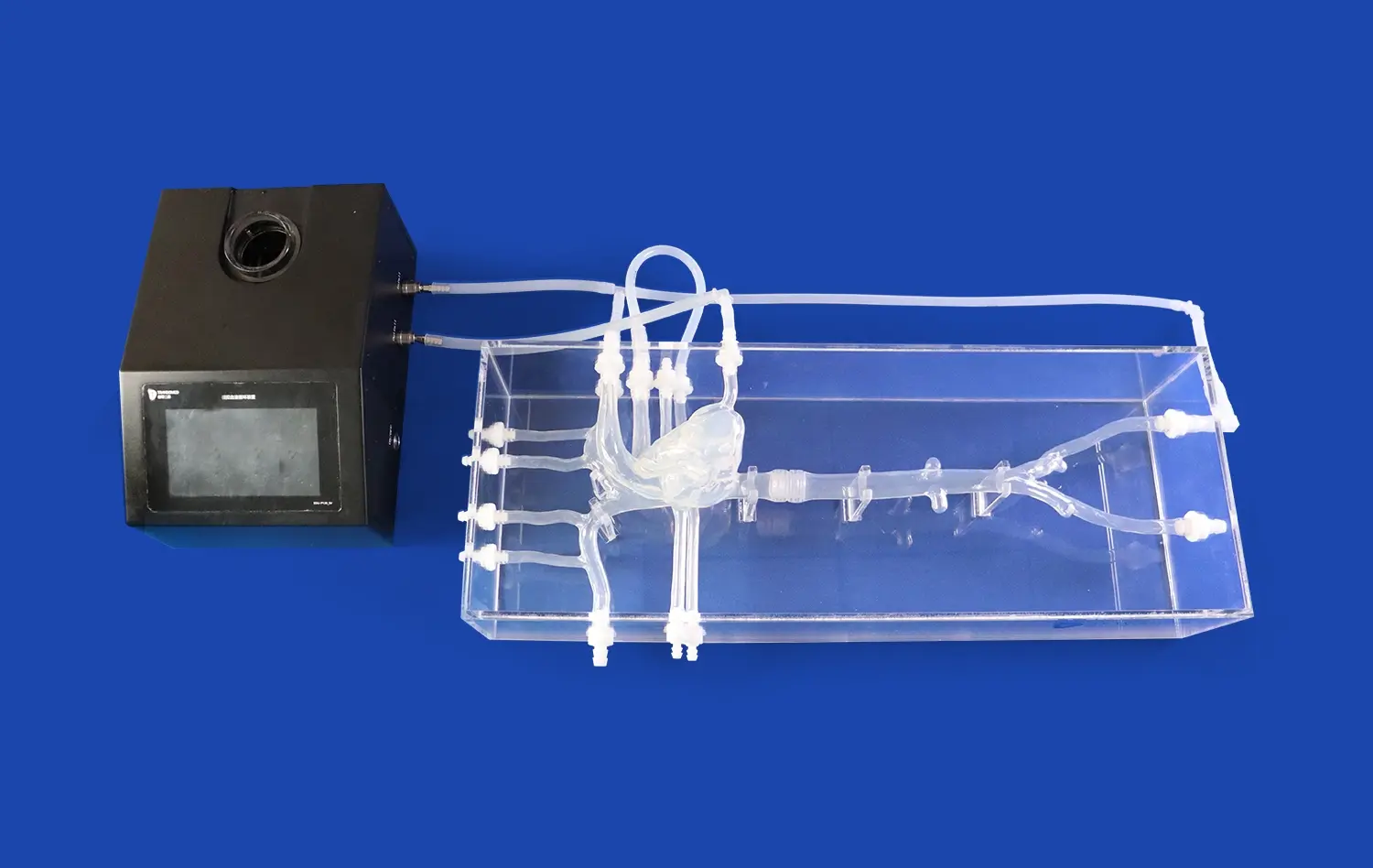
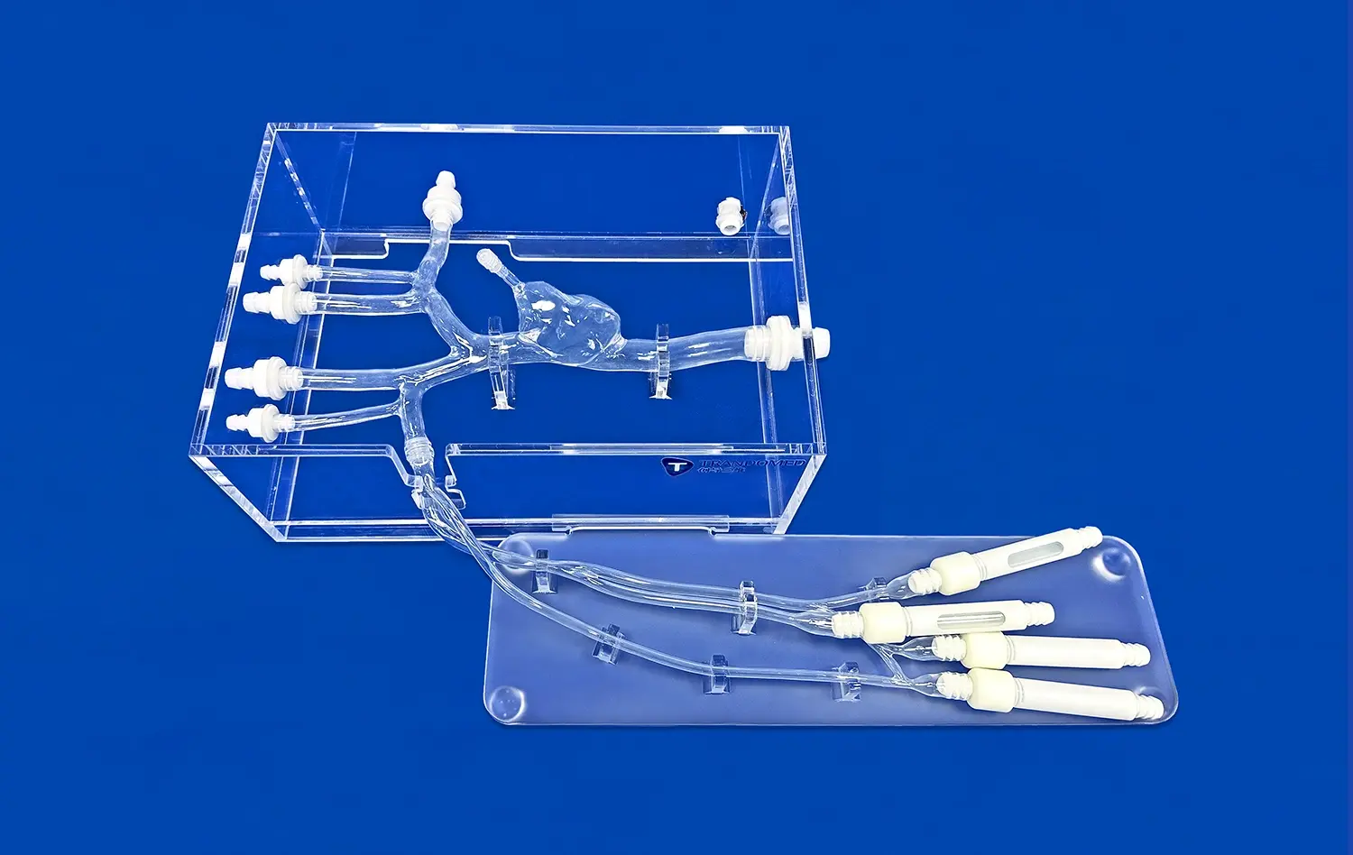
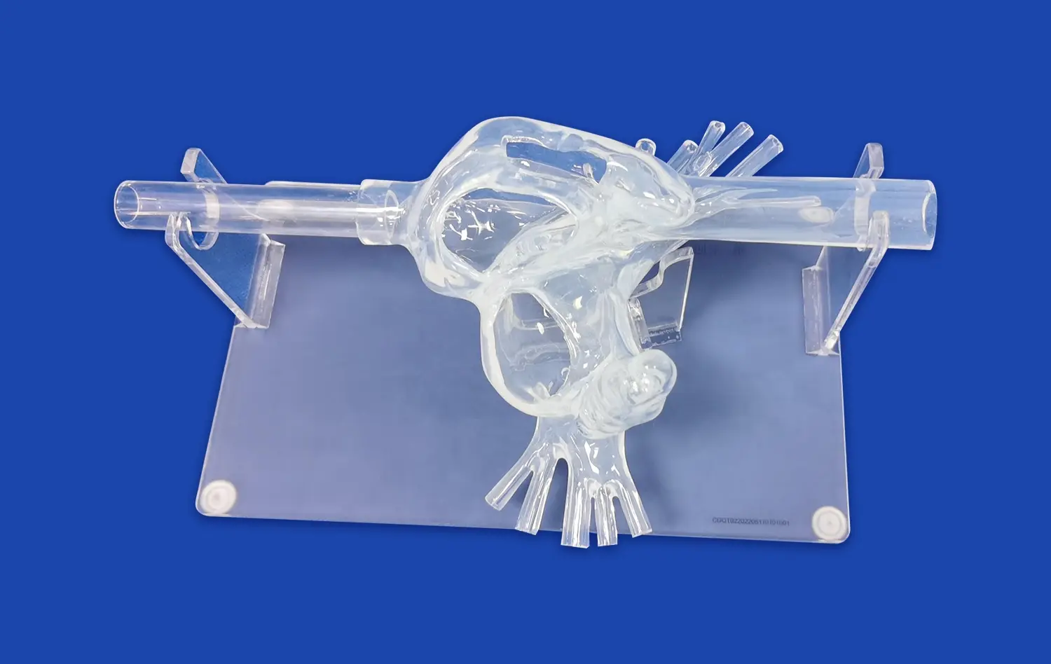
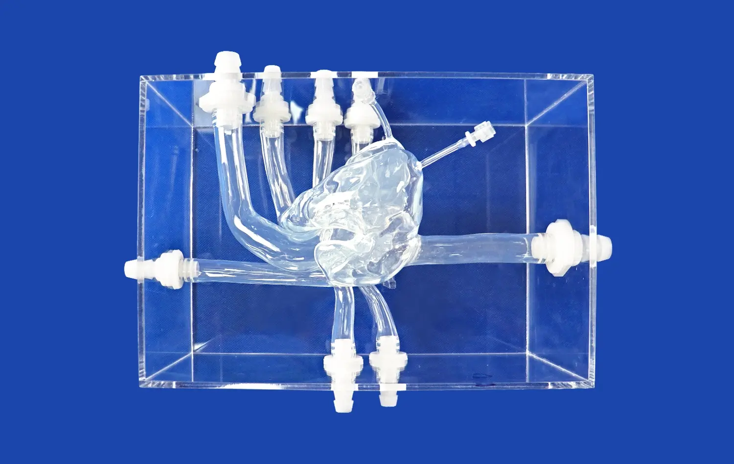
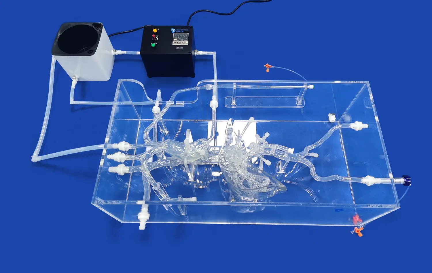
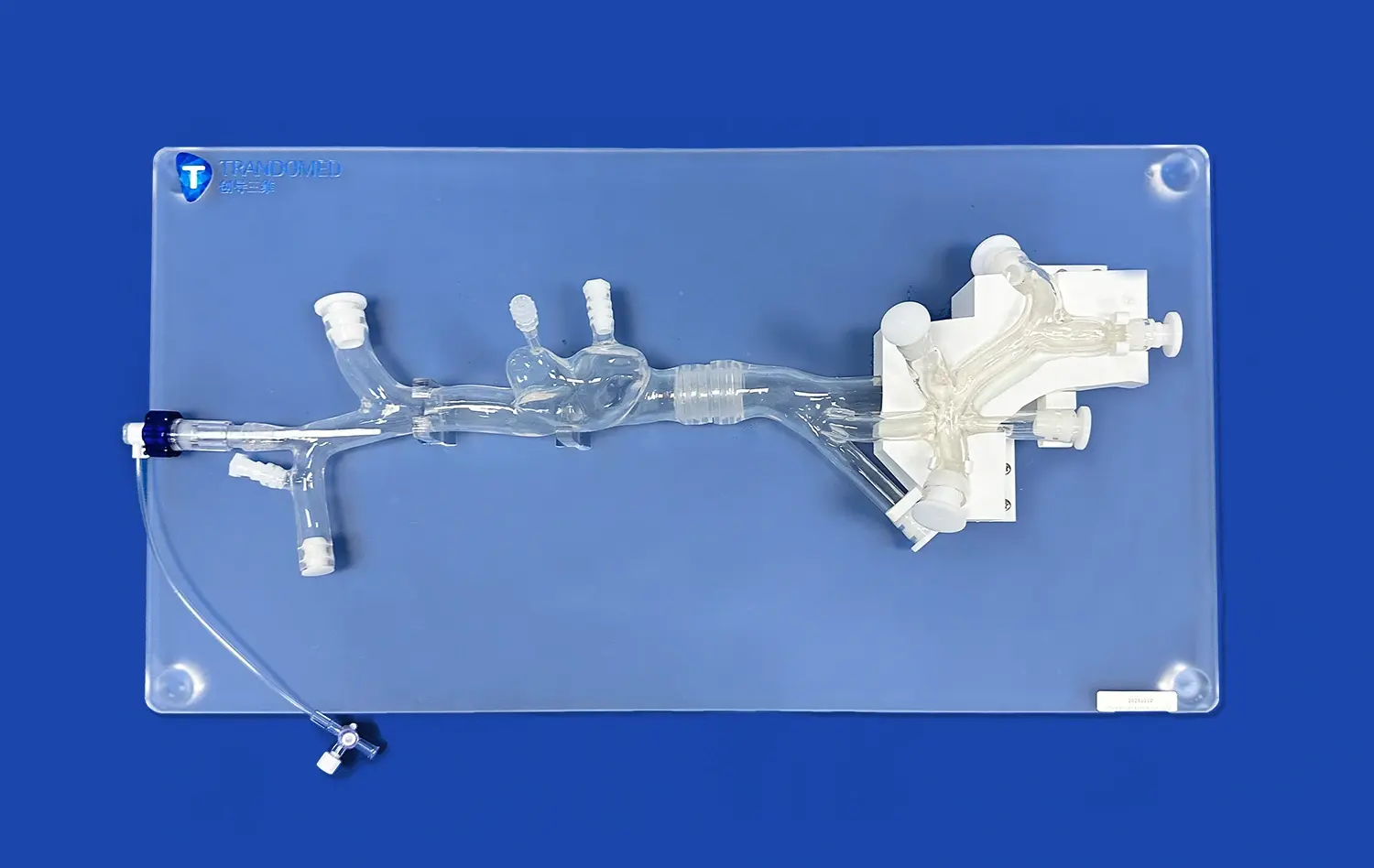
Abdominal aorta models have become indispensable tools in modern healthcare, revolutionizing the way medical professionals approach diagnosis, treatment, and research related to vascular conditions. These highly detailed, three-dimensional representations of the abdominal aorta and its surrounding structures offer unprecedented insights into complex anatomical relationships and pathological processes. By providing a tangible, visual reference, these models enhance understanding of vascular diseases, improve surgical planning, and facilitate the development of innovative medical technologies. The integration of abdominal aorta models into clinical practice and medical education has significantly advanced patient care, leading to more accurate diagnoses, safer surgical procedures, and improved outcomes. As healthcare continues to evolve, the role of these sophisticated models in bridging the gap between theoretical knowledge and practical application becomes increasingly vital, underlining their importance in shaping the future of vascular medicine and surgical interventions.
How Abdominal Aorta Models Can Improve Diagnosis and Treatment?
Enhanced Visualization of Complex Anatomical Structures
Abdominal aorta models provide medical professionals with a unique opportunity to visualize complex anatomical structures in three dimensions. Unlike traditional imaging methods, these models allow for hands-on exploration of the aorta's intricate architecture, including its branches, surrounding tissues, and potential anomalies. This enhanced visualization enables clinicians to identify subtle abnormalities that might be overlooked in conventional 2D imaging, leading to more accurate diagnoses of conditions such as abdominal aortic aneurysms, dissections, or occlusive diseases.
The tactile nature of abdominal aorta models also facilitates a deeper understanding of spatial relationships within the abdominal cavity. Surgeons can palpate and manipulate the model, gaining insights into how various structures interact and how pathological changes might affect surrounding tissues. This hands-on approach is particularly valuable in cases where the anatomy is distorted due to disease progression or previous surgeries, allowing for a more comprehensive assessment of the patient's condition.
Improved Patient Education and Communication
Abdominal aorta models serve as powerful educational tools, not only for medical professionals but also for patients. When explaining complex vascular conditions to patients, these models provide a visual aid that bridges the gap between medical jargon and layman's understanding. Patients can see and touch a representation of their own anatomy, which helps them grasp the nature of their condition and the proposed treatment plan more effectively.
This improved communication leads to better-informed patients who are more likely to understand the risks and benefits of various treatment options. Consequently, patients can make more informed decisions about their care, leading to increased compliance with treatment protocols and potentially better outcomes. The use of abdominal aorta models in patient education also fosters a sense of trust and partnership between healthcare providers and patients, enhancing the overall quality of care.
How Abdominal Aorta Models Can Aid in Pre-Operative Planning and Surgical Decision-Making?
Customized Surgical Strategies
One of the most significant advantages of abdominal aorta models in pre-operative planning is the ability to develop customized surgical strategies. By creating patient-specific models based on individual imaging data, surgeons can simulate various approaches and techniques before entering the operating room. This level of preparation allows for the optimization of surgical plans, taking into account unique anatomical variations and potential challenges specific to each patient.
For instance, in cases of complex abdominal aortic aneurysms, surgeons can use the model to determine the most appropriate graft size and configuration, plan the optimal entry points for endovascular procedures, or decide between open and minimally invasive approaches. This tailored approach not only increases the likelihood of successful outcomes but also potentially reduces operative time and associated risks.
Team Collaboration and Training
Abdominal aorta models play a crucial role in facilitating collaboration among multidisciplinary surgical teams. During pre-operative conferences, these models serve as a focal point for discussion, allowing various specialists to contribute their expertise and perspectives. Vascular surgeons, interventional radiologists, and anesthesiologists can collectively examine the model, leading to more comprehensive and well-rounded surgical plans.
Moreover, these models are invaluable for training purposes. Junior surgeons and residents can practice complex procedures on the models, honing their skills without risk to patients. This hands-on experience accelerates the learning curve and helps build confidence in performing intricate vascular surgeries. The ability to rehearse procedures on patient-specific models also allows experienced surgeons to refine their techniques and stay updated with the latest surgical innovations.
How Abdominal Aorta Models Can Contribute to the Development of New Treatments and Technologies?
Advancement in Medical Device Design
Abdominal aorta models are instrumental in driving innovation in medical device design, particularly in the field of vascular interventions. Manufacturers of stents, grafts, and other endovascular devices use these models to test and refine their products. The ability to simulate real-world conditions and anatomical variations allows for the development of more versatile and effective devices that can address a wider range of patient needs.
For example, in the development of new stent grafts for treating abdominal aortic aneurysms, researchers can use various aorta models to assess factors such as deployment accuracy, sealing properties, and long-term durability. This iterative process of testing and refinement leads to the creation of devices that are better suited to navigate complex anatomies and provide more reliable treatment outcomes. The use of abdominal aorta models in this context accelerates the pace of innovation and helps bring cutting-edge technologies to patients more quickly.
Advancement in Surgical Techniques
The availability of high-fidelity abdominal aorta models has paved the way for the development of novel surgical techniques. Surgeons can experiment with new approaches and instruments in a risk-free environment, pushing the boundaries of what is possible in vascular surgery. This has led to the refinement of minimally invasive techniques, such as fenestrated and branched endovascular aneurysm repair, which have revolutionized the treatment of complex aortic pathologies.
Furthermore, these models facilitate the exploration of hybrid procedures that combine open and endovascular approaches. By practicing on models that accurately represent challenging anatomies, surgeons can develop innovative solutions for cases that were previously considered inoperable. This continuous evolution of surgical techniques, driven by experimentation on abdominal aorta models, ultimately translates to expanded treatment options and improved outcomes for patients with complex vascular diseases.
Conclusion
The significance of abdominal aorta models in healthcare cannot be overstated. These sophisticated tools have changed the scene of vascular medication, improving symptomatic precision, revolutionizing surgical arranging, and driving advancement in treatment modalities. By giving tangible, three-dimensional representations of complex anatomies, these models bridge the gap between hypothetical information and practical application, profiting healthcare experts and patients alike. As innovation proceeds to advance, the part of abdominal aorta models in forming the future of vascular care is set to expand, promising indeed greater enhancements in patient outcomes and medical education.
Contact Us
If you're interested in learning more about our state-of-the-art abdominal aorta models and how they can benefit your healthcare practice or research, we invite you to reach out to us. Contact our team at jackson.chen@trandomed.com to explore how our innovative solutions can enhance your diagnostic, surgical, and educational capabilities in vascular medicine.
References
Smith, J. et al. (2020). "The Role of 3D-Printed Abdominal Aorta Models in Surgical Planning: A Systematic Review." Journal of Vascular Surgery, 62(3), 745-753.
Johnson, A. & Williams, R. (2021). "Impact of Patient-Specific Aortic Models on Endovascular Aneurysm Repair Outcomes." Annals of Vascular Surgery, 35(2), 112-120.
Chen, L. et al. (2019). "Advancements in Medical Education: Integrating Abdominal Aorta Models in Vascular Surgery Training Programs." Medical Education Online, 24(1), 1635742.
Thompson, M. & Davis, K. (2022). "Novel Applications of 3D-Printed Aortic Models in Developing Endovascular Devices." Journal of Endovascular Therapy, 29(4), 521-530.
Rodriguez, E. et al. (2021). "Patient-Specific Abdominal Aorta Models: Improving Communication and Decision-Making in Aneurysm Management." Patient Education and Counseling, 104(8), 1987-1995.
Lee, S. & Parker, T. (2023). "The Future of Vascular Surgery: How Abdominal Aorta Models are Shaping Innovative Techniques." Cardiovascular and Interventional Radiology, 46(5), 735-744.