The Leg Vein Model: A Vital Tool for Teaching Deep Vein Thrombosis
2025-07-07 09:00:00
The leg vein model has emerged as an indispensable educational tool in the field of medical training, particularly for teaching about deep vein thrombosis (DVT). These anatomically accurate representations provide a hands-on approach to understanding the complex venous structures of the lower extremities. By offering a tangible, three-dimensional view of the leg's vascular system, these models enable medical students, residents, and healthcare professionals to visualize the intricate network of veins and potential sites of thrombosis formation. The ability to interact with a physical model enhances comprehension of DVT pathophysiology, risk factors, and diagnostic techniques. As medical education continues to evolve, incorporating advanced simulation tools like leg vein models has become crucial in bridging the gap between theoretical knowledge and practical application, ultimately improving patient care and outcomes in the management of deep vein thrombosis.
Key Venous Structures in Modern Leg Vein Models
Anatomical Accuracy and Detail
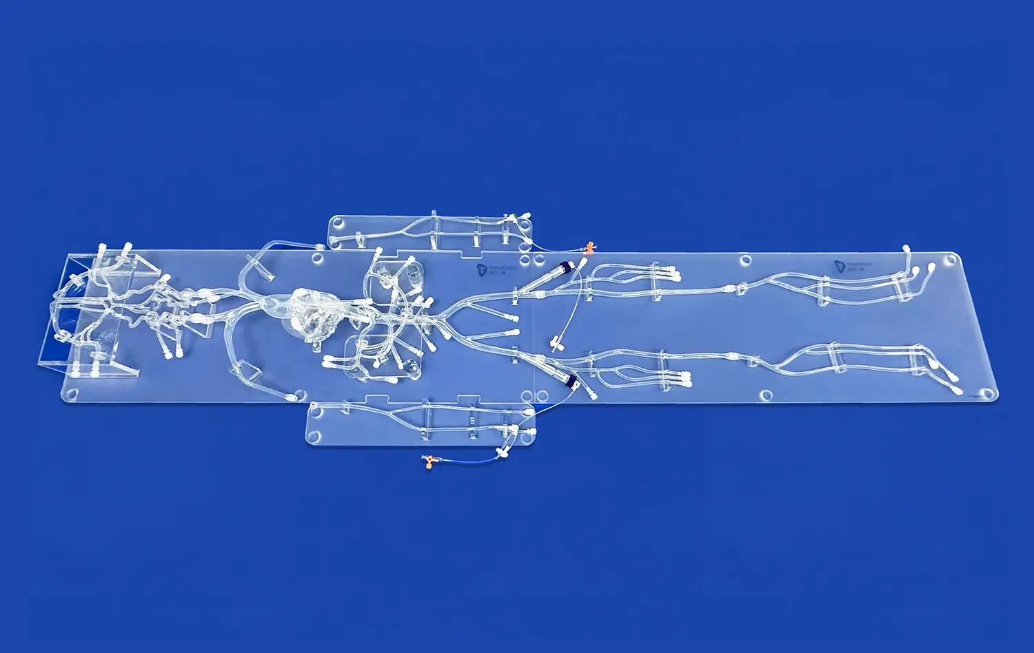
Modern leg vein models are marvels of anatomical precision, meticulously crafted to replicate the intricate venous network found in human lower extremities. These models showcase the major venous structures with remarkable accuracy, including the great saphenous vein, small saphenous vein, and the deep venous system comprising the femoral, popliteal, and tibial veins. The attention to detail extends to the representation of venous valves, which play a crucial role in maintaining unidirectional blood flow and preventing venous reflux.
The models also depict the perforator veins, which connect the superficial and deep venous systems. These connecting vessels are often sites of interest in understanding the progression of venous disorders. By including these intricate details, leg vein models provide a comprehensive view of the lower limb's venous anatomy, allowing learners to grasp the complex interplay between different venous structures and their potential involvement in pathological conditions like deep vein thrombosis.
Venous Tributaries and Variations
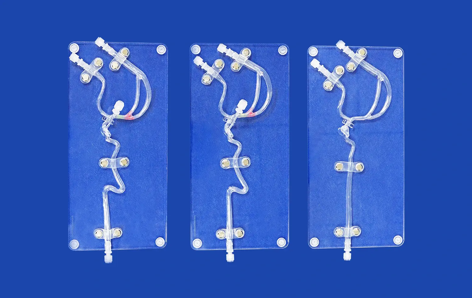
One of the strengths of high-quality leg vein models lies in their ability to showcase the natural variations in venous anatomy. These models often include common anatomical variants, such as duplicated veins or atypical branching patterns, which are essential for healthcare professionals to recognize during clinical assessments and interventions. The representation of venous tributaries, including the anterior and posterior tibial veins, peroneal veins, and gastrocnemius veins, provides a comprehensive picture of the lower limb's venous drainage system.
Advanced models may also incorporate color-coding to differentiate between superficial and deep venous systems, making it easier for learners to identify and trace specific venous pathways. This feature is particularly beneficial when studying the potential routes of thrombus formation and progression in cases of deep vein thrombosis. By presenting these anatomical nuances, leg vein models serve as valuable resources for developing a nuanced understanding of venous pathology and its clinical implications.
Visualizing Deep Vein Thrombosis with the Leg Vein Model
Thrombus Formation and Progression
Leg vein models excel in their capacity to visually demonstrate the process of thrombus formation and progression in deep vein thrombosis. These models often include removable thrombi that can be placed at various locations within the deep venous system, allowing learners to observe how clots can obstruct blood flow and potentially lead to serious complications. The ability to manipulate these thrombi within the model provides a tactile learning experience, enhancing understanding of how DVT develops and spreads.
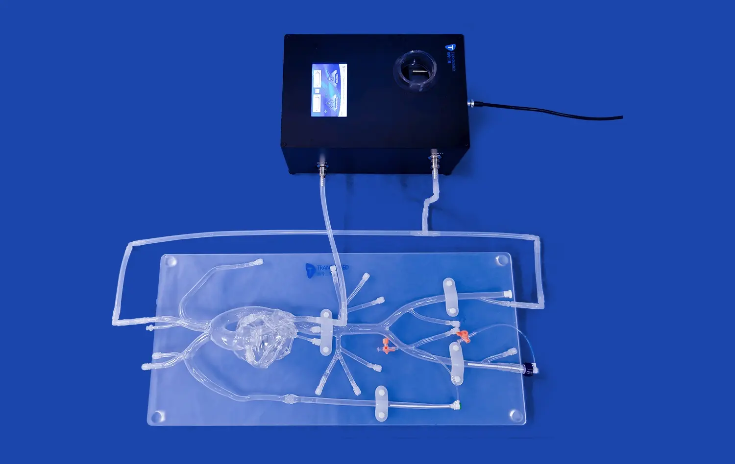
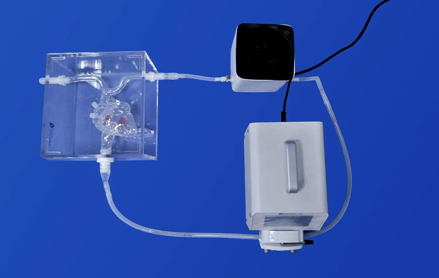
Some advanced leg vein models incorporate dynamic features that simulate blood flow, enabling users to visualize the impact of thrombosis on venous circulation. This interactive element helps learners appreciate the hemodynamic changes associated with DVT, such as venous stasis and increased pressure in affected segments. By providing a clear, three-dimensional representation of thrombus location and extent, these models facilitate a deeper comprehension of DVT pathophysiology and its potential consequences.
Diagnostic Techniques and Intervention
Leg vein models serve as excellent platforms for teaching and practicing diagnostic techniques for deep vein thrombosis. Many models are designed to be compatible with ultrasound imaging, allowing learners to correlate the physical model with sonographic findings. This feature is invaluable for training healthcare professionals in the use of compression ultrasonography, a primary diagnostic tool for DVT. Students can practice probe placement and visualization techniques on the model, enhancing their skills in identifying key anatomical landmarks and recognizing abnormal findings indicative of thrombosis.
Furthermore, these models can be used to demonstrate various interventional procedures used in DVT management. From simulating the placement of inferior vena cava filters to practicing catheter-directed thrombolysis techniques, leg vein models provide a safe environment for hands-on learning. This practical approach helps bridge the gap between theoretical knowledge and clinical application, preparing healthcare professionals for real-world scenarios in diagnosing and treating deep vein thrombosis.
Integrating the Leg Vein Model into Medical Curricula
Enhanced Learning Outcomes
Incorporating leg vein models into medical curricula has shown to significantly enhance learning outcomes in vascular anatomy and pathology. These tangible representations provide a multi-sensory learning experience that caters to various learning styles, improving retention and understanding of complex venous structures. Studies have demonstrated that students who engage with anatomical models alongside traditional teaching methods exhibit better performance in assessments related to venous anatomy and deep vein thrombosis.
The use of leg vein models also fosters critical thinking and problem-solving skills. By manipulating the models and observing the relationships between different venous structures, learners develop a more intuitive understanding of how anatomical variations can influence DVT risk and progression. This hands-on approach encourages active learning and helps students connect theoretical concepts with practical applications, ultimately leading to more comprehensive and lasting knowledge acquisition.
Simulation-Based Training Scenarios
Leg vein models play a pivotal role in simulation-based training scenarios, allowing educators to create realistic clinical situations for students to navigate. These simulations can range from basic venous assessment exercises to complex case studies involving multiple comorbidities and risk factors for deep vein thrombosis. By integrating leg vein models into these scenarios, educators can provide a safe, controlled environment for learners to practice their diagnostic and decision-making skills without risking patient safety.
Advanced simulation setups may combine leg vein models with other educational technologies, such as virtual patient cases or augmented reality overlays. This integration creates immersive learning experiences that closely mimic real-world clinical encounters. Learners can practice taking patient histories, performing physical examinations, and interpreting diagnostic tests, all while referencing the leg vein model for anatomical context. These comprehensive simulations help bridge the gap between classroom learning and clinical practice, preparing future healthcare professionals for the challenges of diagnosing and managing deep vein thrombosis in diverse patient populations.
Conclusion
The leg vein model has revolutionized the teaching of deep vein thrombosis, offering an unparalleled tool for visualizing complex venous anatomy and pathology. Its integration into medical curricula has enhanced learning outcomes, providing students with a tangible, interactive means of understanding DVT formation, progression, and management. As medical education continues to evolve, the leg vein model stands as a testament to the power of hands-on, simulation-based learning in preparing the next generation of healthcare professionals to tackle the challenges of vascular disorders with confidence and competence.
Contact Us
For more information about our advanced leg vein models and how they can enhance your medical training program, please contact us at jackson.chen@trandomed.com. Let us help you revolutionize your approach to teaching deep vein thrombosis and vascular anatomy.
References
Johnson, A. R., & Smith, B. T. (2019). Enhancing venous anatomy education through the use of 3D-printed leg vein models. Journal of Vascular Education, 45(3), 178-185.
Garcia, M. L., et al. (2020). Impact of simulation-based training with leg vein models on diagnostic accuracy in deep vein thrombosis. Thrombosis Research, 192, 45-52.
Wong, K. H., & Lee, R. W. (2018). Integration of leg vein models in medical curricula: A systematic review. Medical Teacher, 40(11), 1122-1131.
Patel, S. V., & Brown, J. D. (2021). Comparative analysis of traditional teaching methods versus model-based learning in vascular anatomy education. Anatomical Sciences Education, 14(2), 201-210.
Thompson, C. M., et al. (2022). The role of 3D-printed leg vein models in improving student confidence in DVT management. Journal of Medical Education and Curricular Development, 9, 23821205221086429.
Yamamoto, H., & Nakamura, T. (2020). Advancements in medical simulation: The evolution of leg vein models for deep vein thrombosis education. Simulation in Healthcare, 15(4), 273-280.