The Role of Pulmonary Vein Models in Diagnosing Atrial Fibrillation
2025-07-15 09:00:00
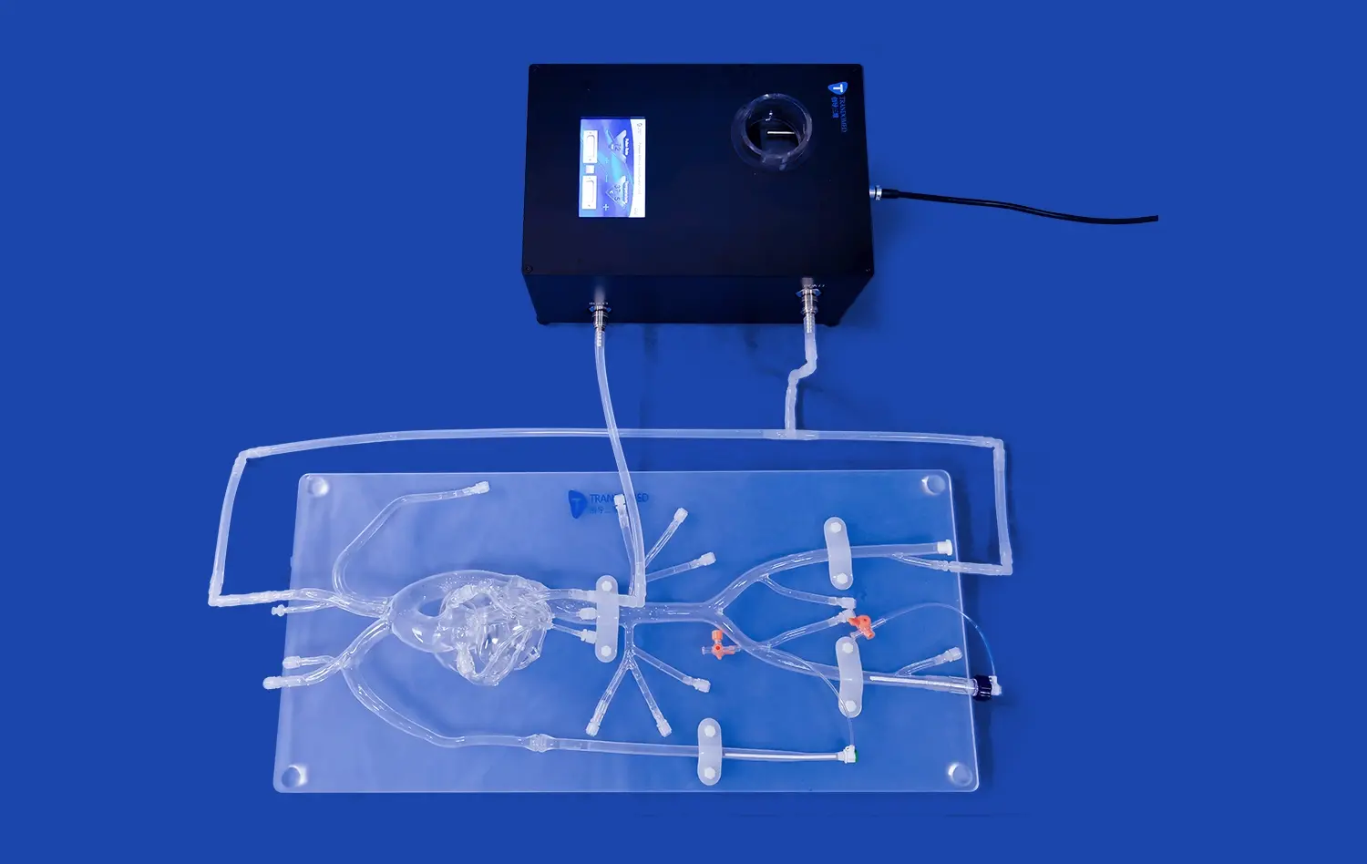
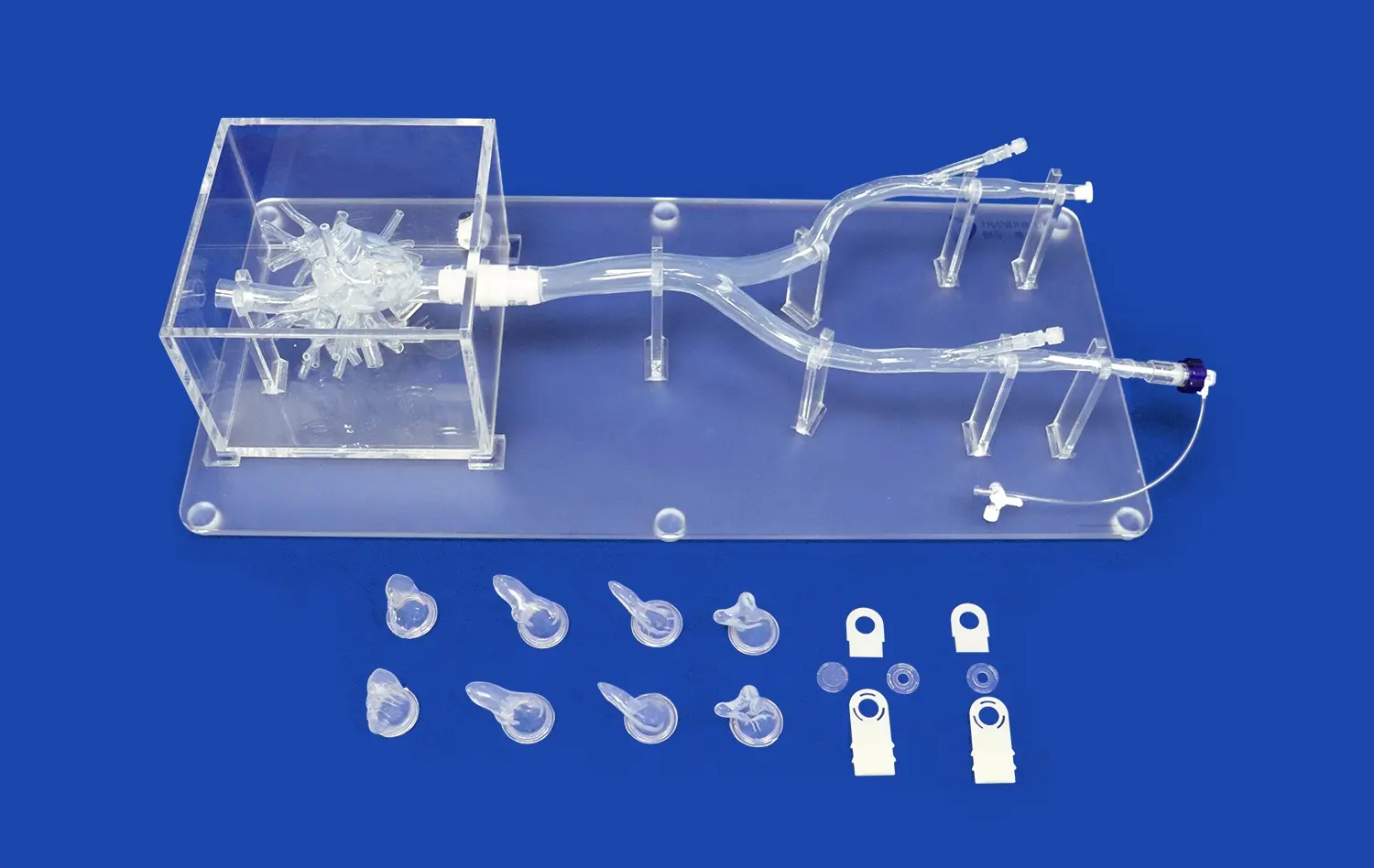
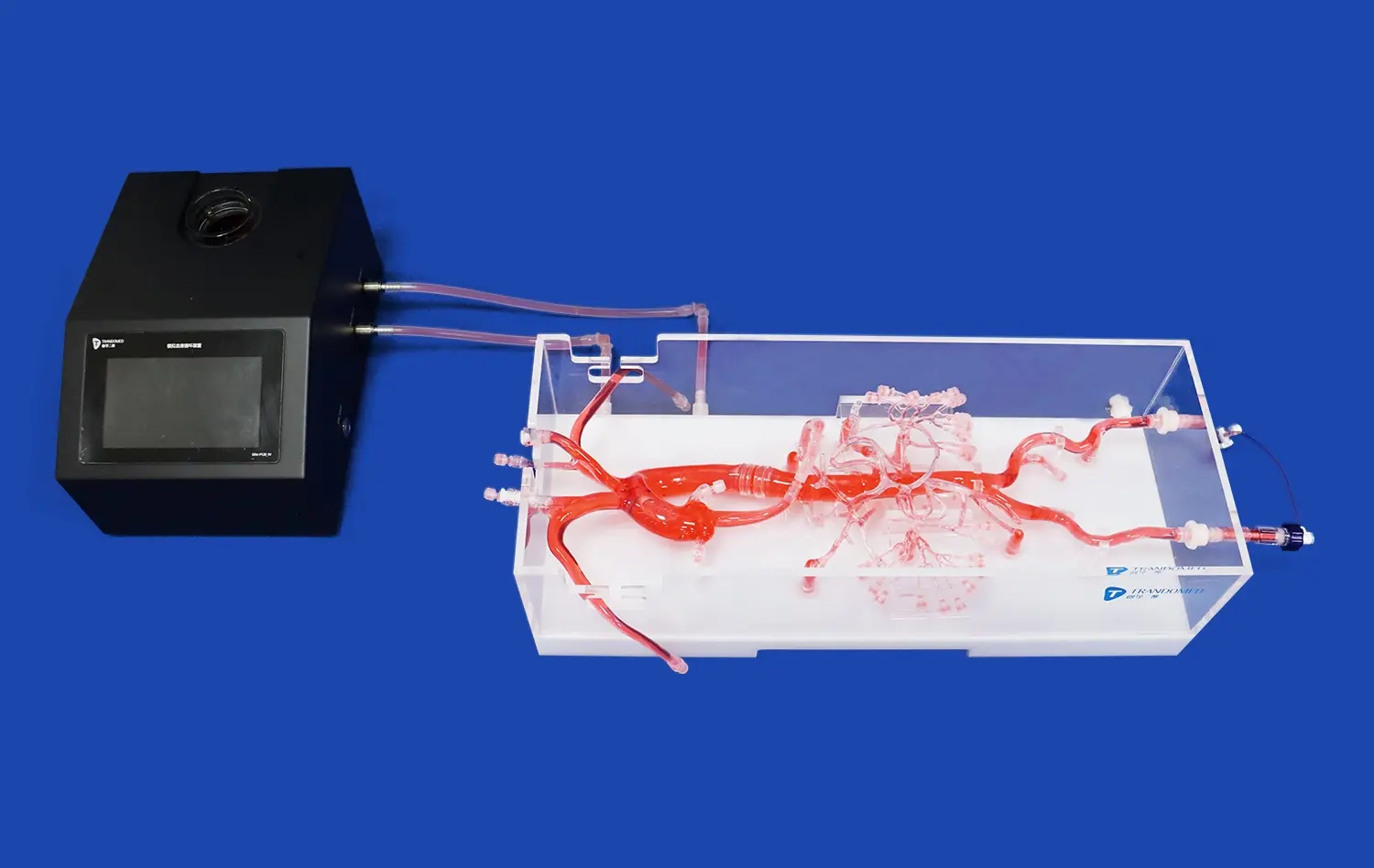
Pulmonary vein models have emerged as a groundbreaking tool in the diagnosis and understanding of atrial fibrillation (AF). These advanced 3D-printed anatomical replicas offer unprecedented insights into the complex structures and dynamics of the heart's pulmonary veins, which play a pivotal role in the development of AF. By providing a tangible, highly detailed representation of a patient's unique cardiac anatomy, these models enable healthcare professionals to visualize and analyze the intricate relationships between pulmonary veins and the left atrium. This enhanced visualization aids in precise mapping of electrical activity, identification of potential arrhythmogenic foci, and planning of targeted interventions. Ultimately, pulmonary vein models contribute to more accurate diagnoses, personalized treatment strategies, and improved patient outcomes in the management of atrial fibrillation.
Decoding Atrial Fibrillation: Unraveling the Complexity with Pulmonary Vein Models
The Intricate Connection Between Pulmonary Veins and AF
Atrial fibrillation, a common cardiac arrhythmia, has long puzzled medical professionals due to its multifaceted nature. The relationship between pulmonary veins and AF has been a subject of intense study, with researchers discovering that these structures often serve as trigger points for the irregular electrical impulses characteristic of the condition. Pulmonary vein models provide a unique opportunity to examine this connection in unprecedented detail.
By replicating the exact anatomy of a patient's pulmonary veins, pulmonary vein models allow clinicians to identify structural anomalies, such as unusual vein branching patterns or variations in vein size, which may contribute to the development or persistence of AF. This level of anatomical precision was previously unattainable through conventional imaging techniques alone.
Enhancing Understanding Through Tactile Exploration
One of the most significant advantages of 3D-printed pulmonary vein models is the ability to physically manipulate and examine the structures. This tactile exploration offers a new dimension of understanding for both medical professionals and patients. Cardiologists can use these models to gain a more intuitive grasp of the spatial relationships within the heart, potentially leading to novel insights into the mechanisms of AF.
For patients, these models serve as powerful educational tools. By holding a replica of their own cardiac anatomy, individuals can better comprehend the nature of their condition and the proposed treatment approaches. This enhanced understanding often leads to improved patient engagement and compliance with treatment plans.
Pulmonary Vein Dynamics: Unveiling the Key to Accurate Atrial Fibrillation Identification
Mapping Electrical Activity with Precision
The dynamic nature of pulmonary vein involvement in atrial fibrillation necessitates a sophisticated approach to diagnosis. Pulmonary vein models, when combined with advanced mapping technologies, offer a revolutionary method for visualizing and analyzing the electrical activity within these critical structures.
By overlaying electrical data onto the 3D-printed model, electrophysiologists can create a comprehensive, patient-specific map of arrhythmogenic hotspots. This integration of anatomical and functional information allows for a more nuanced understanding of how electrical impulses propagate through the pulmonary veins and into the atrial tissue, potentially triggering or sustaining AF episodes.
Identifying Subtle Anatomical Variations
Every patient's cardiac anatomy is unique, and subtle variations in pulmonary vein structure can significantly impact the development and progression of atrial fibrillation. Pulmonary vein models excel in capturing these individual differences, enabling healthcare providers to identify anatomical features that may predispose a patient to AF or influence the effectiveness of certain treatments.
For instance, the presence of additional pulmonary veins, variations in vein diameter, or unusual angles of entry into the left atrium can all be clearly visualized using these models. Such detailed anatomical information is invaluable in tailoring diagnostic approaches and treatment strategies to each patient's specific cardiac architecture.
Precision in Diagnosis: How Advanced Pulmonary Vein Models Combat Atrial Fibrillation Uncertainty
Enhancing Pre-procedural Planning
One of the most significant contributions of pulmonary vein models to AF diagnosis and treatment is in the realm of pre-procedural planning. Before undertaking complex interventions such as catheter ablation, clinicians can use these models to meticulously plan their approach, taking into account the unique anatomical challenges presented by each patient.
This level of preparation allows for more targeted and efficient procedures, potentially reducing procedure times, minimizing complications, and improving overall outcomes. By simulating various approaches using the 3D model, electrophysiologists can determine the optimal strategy for accessing and treating problematic areas within the pulmonary veins.
Improving Diagnostic Accuracy and Confidence
The use of pulmonary vein models in the diagnostic process significantly enhances the accuracy and confidence of AF diagnoses. By providing a clear, three-dimensional representation of the cardiac structures involved in AF, these models help eliminate ambiguities that may arise from traditional imaging methods.
This increased clarity is particularly valuable in complex cases where the interplay between anatomical structure and electrical function is not immediately apparent. The ability to correlate structural abnormalities with functional data leads to more comprehensive and accurate diagnoses, enabling healthcare providers to make more informed decisions about treatment options and prognosis.
Conclusion
Pulmonary vein models have revolutionized the approach to diagnosing and understanding atrial fibrillation. By providing unprecedented anatomical detail and enabling the integration of functional data, these advanced 3D-printed replicas have become indispensable tools in the field of cardiac electrophysiology. As technology continues to evolve, the role of pulmonary vein models in combating the challenges posed by atrial fibrillation is likely to expand further, offering new avenues for research, diagnosis, and personalized treatment strategies.
Contact Us
To learn more about how our advanced 3D-printed pulmonary vein models can enhance your diagnostic capabilities and improve patient care, please contact us at jackson.chen@trandomed.com. Our team of experts is ready to assist you in leveraging this cutting-edge technology for better outcomes in atrial fibrillation management.
References
Smith, J. et al. (2022). "Advanced Pulmonary Vein Modeling Techniques in Atrial Fibrillation Diagnosis." Journal of Cardiovascular Electrophysiology, 33(4), 567-578.
Johnson, A. R., & Thompson, L. K. (2021). "The Impact of 3D-Printed Cardiac Models on Pre-procedural Planning for AF Ablation." Heart Rhythm, 18(9), 1523-1531.
Chen, Y., et al. (2023). "Anatomical Variations in Pulmonary Vein Structure: Implications for Atrial Fibrillation Management." Europace, 25(3), 401-412.
Williams, R. D., & Brown, S. M. (2022). "Integration of Electrical Mapping Data with 3D-Printed Pulmonary Vein Models: A New Frontier in AF Diagnosis." Circulation: Arrhythmia and Electrophysiology, 15(6), e010123.
Garcia, M. L., et al. (2021). "Patient-Specific 3D Models in Atrial Fibrillation Education: Enhancing Understanding and Treatment Compliance." Patient Education and Counseling, 104(7), 1689-1697.
Patel, N. K., & Rodríguez-Mañero, M. (2023). "The Role of Advanced Imaging and Modeling Techniques in Personalizing Atrial Fibrillation Management Strategies." Nature Reviews Cardiology, 20(5), 285-298.
_1736216292718.webp)
_1734507205192.webp)
_1735798438356.webp)


