Why Are Mitral Valve Models Vital in Medical Curriculum?
Enhanced Anatomical Understanding
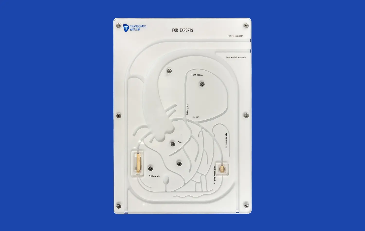
Mitral valve models provide an unparalleled opportunity for students to grasp the intricate anatomy of the heart. Unlike two-dimensional textbook illustrations, these three-dimensional representations allow learners to visualize the spatial relationships between various cardiac structures. The Mitral Valve Model (XXD006) from Trandomed, for instance, offers a comprehensive view from the femoral vein to the right heart, including key vascular structures like the iliac veins and inferior vena cava. This holistic approach enables students to develop a more accurate mental map of cardiac anatomy, crucial for diagnosing and treating mitral valve disorders.
Realistic Simulation of Pathological Conditions
Advanced mitral valve models can simulate various pathological conditions, such as mitral valve prolapse or regurgitation. This feature is invaluable for medical students and residents who may not frequently encounter these conditions in clinical settings. By manipulating these models, learners can observe how structural abnormalities affect valve function, deepening their understanding of disease processes. The ability to replicate different mitral valve conditions allows for a more comprehensive education, preparing future healthcare professionals to recognize and address a wide range of cardiac issues.
Safe Environment for Procedural Practice
One of the most significant advantages of using mitral valve models in medical education is the provision of a safe environment for procedural practice. Aspiring cardiac surgeons can hone their skills without the pressure of operating on actual patients. These models allow for repetitive practice of complex procedures like mitral valve replacement (MVR) or repair techniques. The modular design of some models, such as those offered by Trandomed, even permits the simulation of different valve replacement scenarios, providing a versatile training platform that closely mimics real-life surgical challenges.
Skill Development and Knowledge Retention with Mitral Valve Models
Hands-on Experience in Valve Assessment
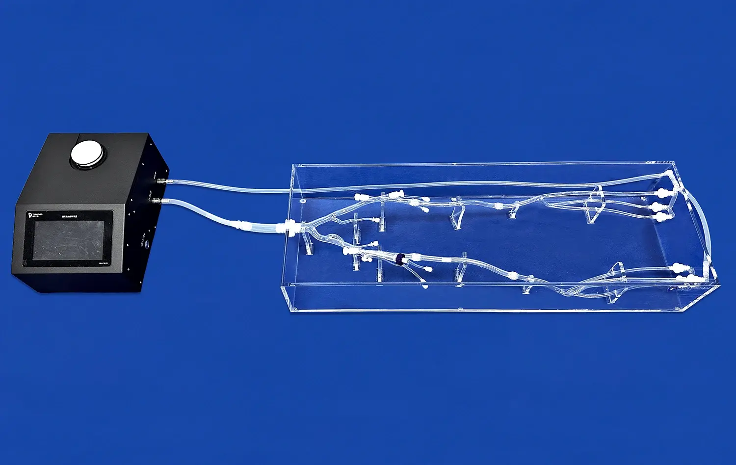
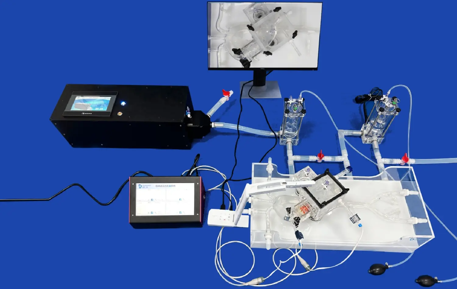
Mitral valve models play a crucial role in developing the tactile skills necessary for valve assessment. Medical students and residents can practice palpating the valve structures, identifying abnormalities, and gauging the severity of conditions like mitral stenosis or regurgitation. This hands-on experience is invaluable in building confidence and competence in physical examination techniques. Moreover, when connected to simulation systems like heart pumps, these models can demonstrate the dynamic function of the mitral valve, allowing learners to observe and understand the opening and closing mechanics in real-time.
Mastering Surgical Techniques
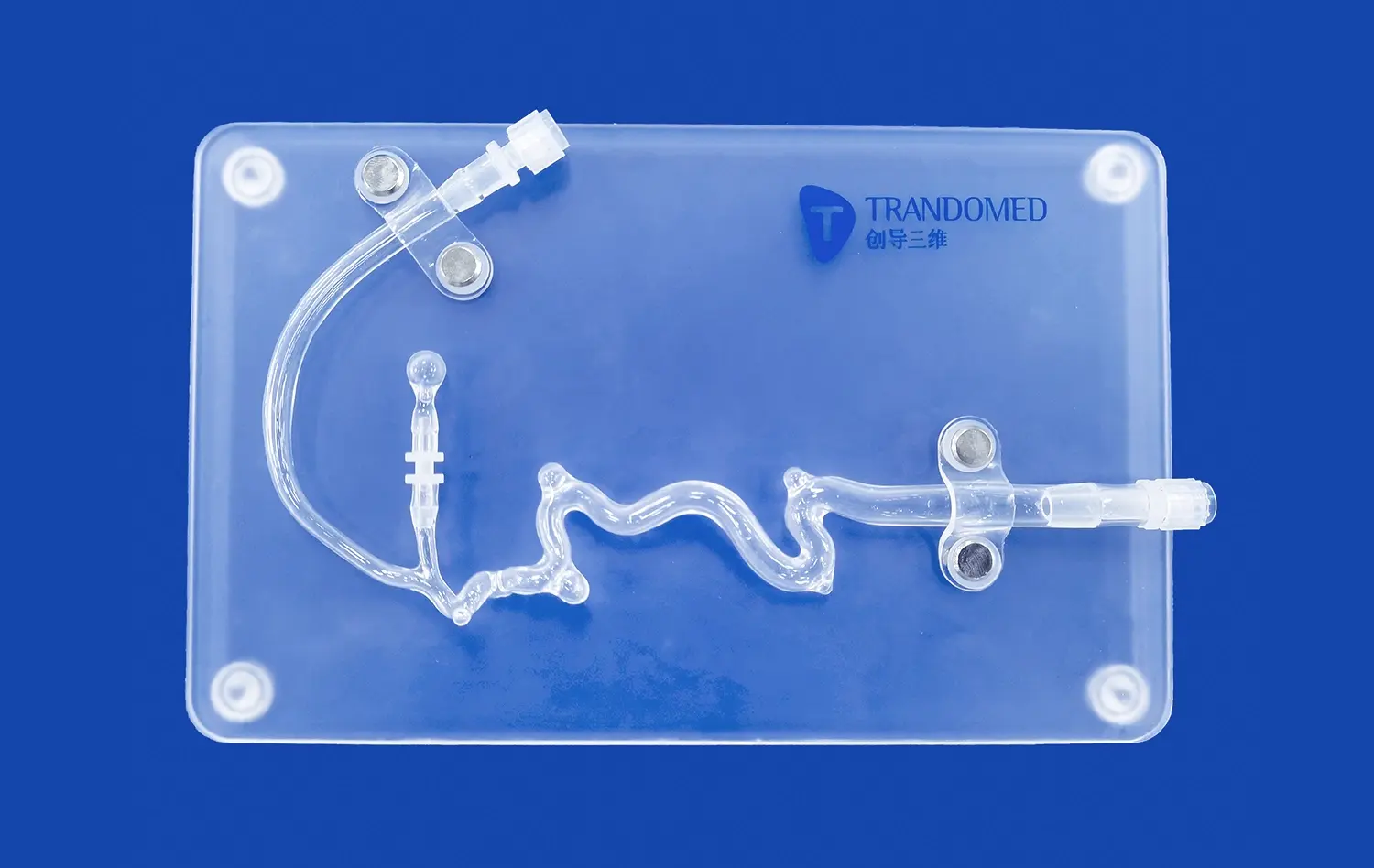
For surgical trainees, mitral valve models offer an unparalleled opportunity to master complex surgical techniques. These models allow for the practice of suturing techniques, valve repair procedures, and the placement of prosthetic valves. The use of high-fidelity silicone materials, such as the Silicone Shore 40A used in Trandomed's models, provides a realistic feel that closely mimics human tissue. This realism is crucial for developing the fine motor skills and spatial awareness required in cardiac surgery. Trainees can practice repeatedly, refining their techniques without the time constraints or risks associated with operating on live patients.
Long-term Retention of Anatomical Knowledge
The use of mitral valve models in medical education significantly enhances long-term retention of anatomical knowledge. The multi-sensory learning experience provided by these models – visual, tactile, and sometimes auditory (in the case of simulated heart sounds) – creates stronger neural connections, leading to better recall of information. This improved retention is particularly beneficial in the field of cardiology, where a thorough understanding of valve anatomy is crucial for accurate diagnosis and treatment planning. Regular interaction with these models throughout the medical curriculum reinforces learning and helps bridge the gap between theoretical knowledge and practical application.
Integration of Mitral Valve Models into Clinical Education Programs
Curriculum Design and Implementation
Integrating mitral valve models into clinical education programs requires thoughtful curriculum design. Medical educators are increasingly recognizing the value of these tools and incorporating them at various stages of training. For instance, in preclinical years, students might use basic models to learn valve anatomy and physiology. As they progress, more advanced models can be introduced to teach pathological conditions and surgical techniques. The modular nature of some mitral valve models, like those offered by Trandomed, allows for a scalable approach to learning, where complexity can be adjusted to match the students' growing knowledge and skills.
Assessment and Competency Evaluation
Mitral valve models serve as excellent tools for assessing student competency in cardiac anatomy and surgical skills. Educators can design objective structured clinical examinations (OSCEs) that incorporate these models, evaluating students' ability to identify structures, diagnose conditions, and perform simulated procedures. This standardized approach to assessment ensures that all students meet the required competency levels before progressing to clinical rotations or surgical training. The use of high-fidelity models in these assessments provides a more accurate representation of a student's readiness for real-world clinical scenarios.
Continuous Professional Development
The benefits of mitral valve models extend beyond undergraduate and postgraduate medical education. These tools play a crucial role in continuous professional development for practicing cardiologists and cardiac surgeons. Advanced models can be used to simulate rare or complex cases, allowing experienced professionals to maintain and upgrade their skills. Workshops and training sessions utilizing these models provide opportunities for surgeons to learn new techniques or practice innovative procedures in a risk-free environment. This ongoing education is essential in a field where technological advancements and surgical techniques are constantly evolving.
Conclusion
The integration of mitral valve models in medical education represents a significant leap forward in preparing healthcare professionals for the complexities of cardiac care. These innovative tools enhance anatomical understanding, provide safe environments for skill development, and offer realistic simulations for both basic and advanced training. As medical education continues to evolve, the role of high-fidelity models like those produced by Trandomed will undoubtedly become even more central to curriculum design and implementation. By bridging the gap between theoretical knowledge and practical application, mitral valve models are not just educational tools; they are catalysts for improved patient care and advancements in cardiovascular medicine.
Contact Us
To explore how Trandomed's cutting-edge mitral valve models can elevate your medical education program, contact us at jackson.chen@trandomed.com. Our team is dedicated to providing customized solutions that meet the unique needs of your institution, ensuring that your students and professionals have access to the most advanced and realistic training tools available in cardiac education.
_1736214519364.webp)
_1734507815464.webp)