Training Neurovascular Procedures with a Circle of Willis Brain Model
2026-01-19 17:50:57
To get good at neurovascular procedures, you need to practice a lot and be able to use modeling tools that are realistic in terms of anatomy. The circle of willis brain model has become an important teaching tool for medical schools, hospitals, and study centers that want to improve their clinical skills. This special anatomical model copies the complicated artery network at the base of the brain. This lets trainees practice delicate procedures like aneurysm coiling, thrombectomy, and catheter guidance without any risk. As simulations become more common in healthcare training, these cerebral models help bridge the gap between academic knowledge and understanding of actual procedures. This improves patient results and lowers professional mistakes.
Understanding the Circle of Willis Brain Model for Neurovascular Training
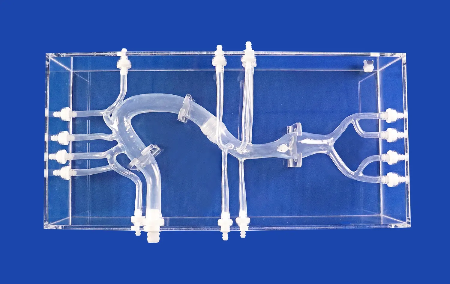
One of the most important arterial systems in the body is the Circle of Willis. This artery ring surrounds the optic chiasm and infundibulum of the pituitary gland and is located on the bottom surface of the brain in the interpeduncular cistern of the subarachnoid space. Its main job is to provide extra blood flow between the front and back parts of the brain, keeping certain areas from going into ischemia when blood vessel disease or damage happens.
Anatomical Components and Functional Significance
For neurovascular training to work, you need to know about the five main parts of this vascular loop. When the anterior cerebral arteries meet at their A1 segments, they join to the anterior communication artery. The internal carotid arteries also contribute at their tips. Here, the posterior cerebral arteries connect with the posterior communicating arteries at their P1 segments to make this protected circulation loop whole. It's interesting that only 20–25% of people have a full, uniform Circle of Willis. The rest have some difference or hypoplasia.
Advanced computer models can copy these differences in anatomy along with common diseases. Some good cerebrovascular models have tumors in common places, like the basilar artery, the ocular section of the carotid artery, and the branches of the middle cerebral artery. They also show stenosis tumors that look like real-life clinical appearances. This helps training identify and treat a range of neurovascular diseases.
Pathological Representations in Training Models
In medical school, students have to learn about diseases and disorders before they can work with patients. Brain circulation models with a high level of detail include accurate images of cerebral aneurysms, intracranial stenosis, and thrombotic occlusions. These features help students see how changes in blood flow affect tissues nearby and comprehend how quickly they need to act to help people who have recently had a stroke.
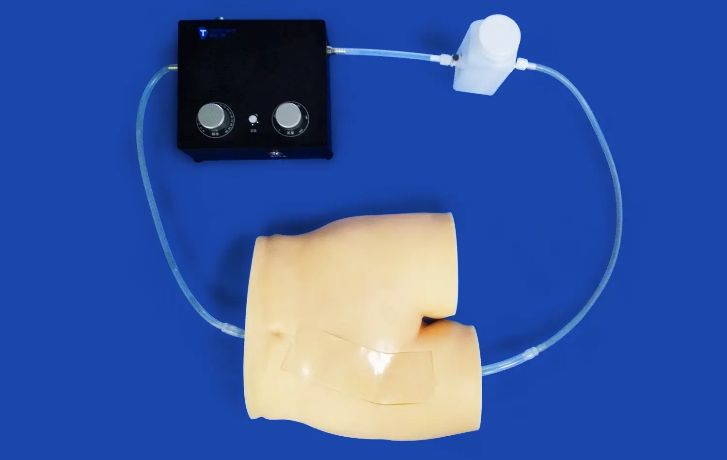
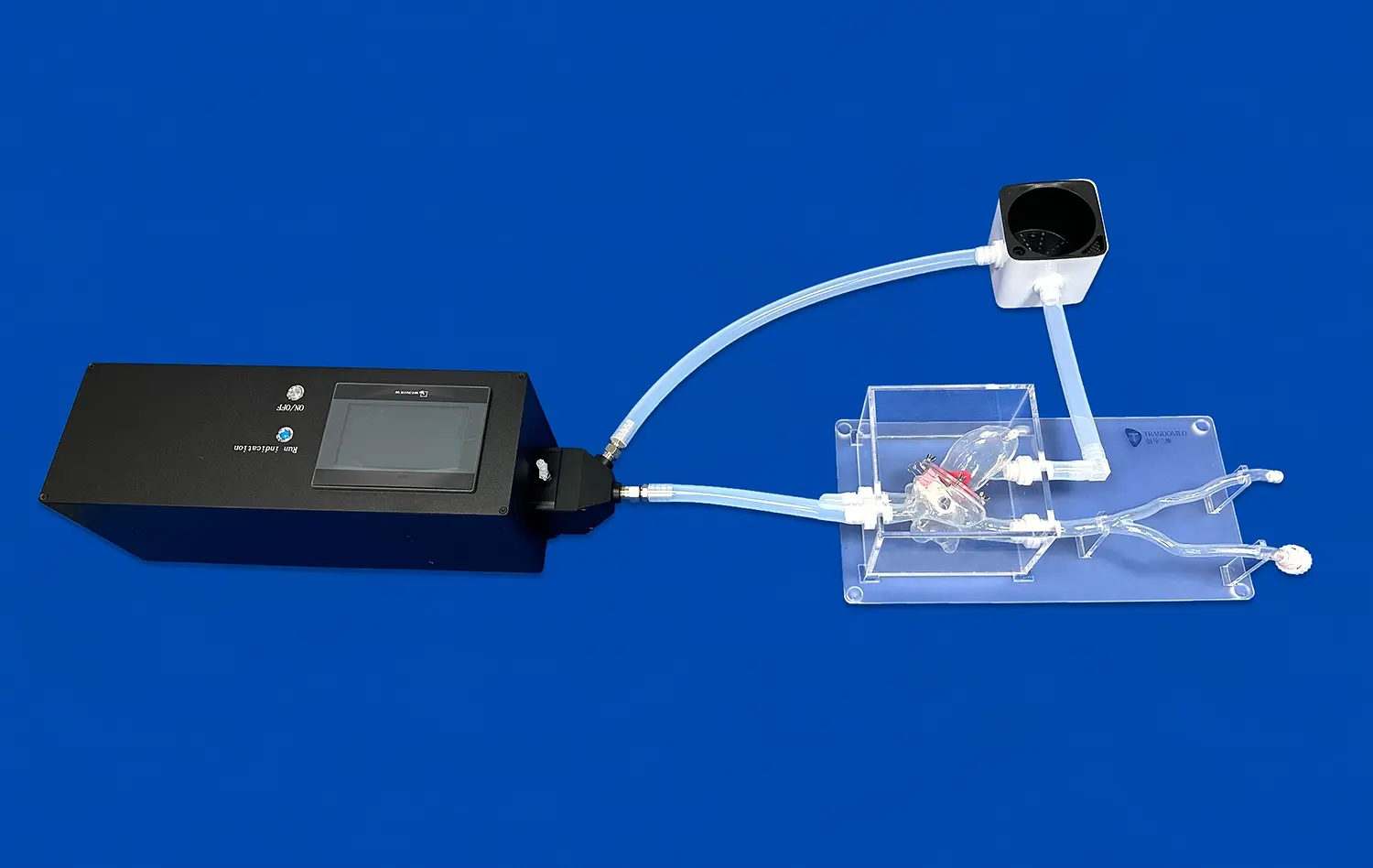
When it comes to high-end models, how they feel is very important. Simulators that imitate the feel and stiffness of real artery walls are helpful for doctors because they give them haptic feedback while they place catheters and deploy devices. This sense factor improves the growth of muscle memory and trust in the procedure, which has a direct effect on success in the operating room.
Selecting the Best Circle of Willis Brain Model for Effective Training
When buying neurovascular modeling tools like the circle of willis brain model, it's important to carefully consider a number of things. On the market, you can find a wide range of choices, from hard plastic anatomical models to high-tech silicone-based procedural teachers. Each of these has its own teaching purpose.
Material Considerations and Durability
The choice of building material has a huge effect on both how realistic it is and how long it lasts. When it comes to feel, silicone-based models, especially those made with Shore 40A durometer silicone, are more realistic than hard plastic options. This type of material closely matches the flexibility and stiffness of human brain vessels, so it can be used for many catheter insertions without breaking down much.
Durability is especially important for places that hold a lot of training classes. Models that are meant to be used a lot should be able to handle hundreds of procedures without losing any of their physical integrity. At popular entry places, reinforced vessel walls stop the walls from breaking too soon. This extends the useful life and lowers the cost of replacement over time.
Customization Capabilities for Specialized Training
B2B buying managers should give more weight to providers that allow customization without charging too much for design services. By letting training programs know about specific learning goals or disease frequency trends in an area, aneurysm number, size, and location can be targeted. Advanced makers can meet requests based on real CT and MRI scans of patients, making models that are unique to each patient for complicated case preparation.
Customization goes beyond how an aneurysm is set up. Models that include a way to change the amount of stenosis at the M1 section of the middle cerebral artery or carotid bifurcation are helpful for training centers that focus on stroke prevention. For research labs to make new arterial devices, they need models that can be changed to fit different conditions, like different branch shapes or altered vessel tortuosity.
Supplier Evaluation Criteria
When buying workers choose a cerebrovascular model maker, they should look at a number of important factors. Technical know-how is very important. Suppliers who use reverse 3D modeling technology from real human image data provide more accurate anatomy than those who use general models. Also, the ways things are made should be looked at closely. For example, 3D printing modeling technology is better at keeping things consistent than standard casting methods.
Standards for certification and quality control for a circle of willis brain model give you an idea of how reliable something is. Well-known companies use strict checking procedures to make sure that every unit meets the required material qualities and size limits. Delivery times are set by global transportation powers. This is especially important for schools that use academic calendars with set training plan due dates.
Integrating the Circle of Willis Brain Model in Neurovascular Procedure Training
For anatomical models to be effectively added to program design, there needs to be organized planning for their use. The change from passively watching to actively taking part in simulations is a turning point in neurovascular education.
Curriculum Integration Strategies
The best results are seen when medical schools and clinical skills centers gradually introduce cerebrovascular models into training routines. The first part of the lesson is all about orienting the body - learning how to find blood vessel branches, understand how blood flows, and tell the difference between standard and unusual anatomy. In later classes, students learn about abnormal traits that make it harder for them to figure out where an aneurysm is and how bad the stenosis is by looking at it and touching it.
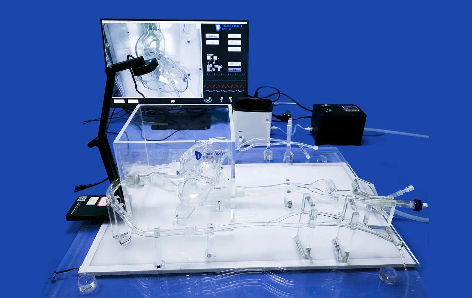
Advanced training modules emphasize procedural skills development. Trainees practice moving a guidewire through blood vessels that are twisted, placing a tube inside an aneurysm sac, and putting in place stents or bands that shift blood flow. The controlled setting lets mistakes happen without hurting the patient, which encourages trying new things and getting better at what you do.
Simulation-Based Competency Assessment
With objective performance measures, simulation training goes from being based on biased observation to a measurable assessment of skill. Training programs time how long it takes to finish a procedure, how accurately the device is placed, and how quickly problems are identified. These tests show where students need to improve their skills before they can move on to guided clinical cases.
Case studies from top neurosurgery residency programs show that trainees' trust and the success rate of procedures go up after they do a lot of computer training. In a published study, residents who did 20 virtual thrombectomy procedures had 40% faster recanalization times during their first three guided patient treatments than their peers who had not done any simulations.
Bridging Theory and Clinical Practice
High-fidelity models are useful for teaching more than just technical skills. Working with accurate models helps students understand how imaging results relate to the three-dimensional structure of the circulatory system. When using fluoroscopic direction to find your way during real treatments, this knowledge of space is very helpful.
Multidisciplinary training situations make people even more ready for clinical work. Interventional neurologists, neurosurgeons, anesthesiologists, and nurse staff work together in simulations that mimic how the operating room really works. This helps the team communicate better and understand their roles better during situations that need to be handled quickly.
Conclusion
Access to physically accurate and functionally realistic modeling tools like the circle of willis brain model is very important for the progress of neurovascular training. High-quality brain models help doctors get better at their jobs by letting them practice in safe settings, which improves patient safety and clinical results. As simulation-based learning becomes more common in healthcare education, buying better training tools is both necessary for the students and a smart move for the school as a whole. When companies put physical correctness, customization options, and source dependability at the top of their list of priorities, they set themselves up to provide excellent clinical education that meets changing professional standards.
FAQs
What makes a circle of willis brain model perfect for training the nerves and blood vessels?
An ideal neurovascular training model is one that is both accurate in terms of anatomy and usefulness. The model should show the anterior cerebral arteries, the internal carotid arteries, the posterior cerebral arteries, and the communication veins correctly in terms of where they are in space. The material's features must match the flexibility of a human blood vessel so that it gives the right physical input when the catheter is being moved. Putting common disease traits like aneurysms and stenosis lesions in places that are practically important makes the learning more useful. Cost-effectiveness over long training times is ensured by the ability to be used over and over again without breaking down.
How can schools make sure that they meet certain training needs?
The first step in compatibility testing is to make sure that training goals are clear. For institutions that teach diagnostic skills, models that focus on recognizing anatomy differences are needed. For procedure training programs, simulations that can hold real invasive devices are needed. Talking to providers about their customization options makes sure that product specs and program goals are in line with each other. By asking for sample units or trial sessions, you can check out the physical features and useful performance before committing to buy a lot of them. Suppliers with experience in medical education usually offer advice on how to match the features of their products with the needs of each school.
What are the usual lead times for large sales, and are there ways to make them unique?
Standard wait times for making well-known cerebrovascular types are between 7 and 10 days for stock setups. Custom changes, like placing an artery in a different place, adjusting the depth of a stenosis, or making structural copies that are unique to a patient, may make the process take longer, based on how complicated the design is. Manufacturers often give priority ordering to bulk orders and set aside extra production capacity for big group purchases. Customization options include changing the number and location of aneurysms, the degree to which vessels are tortuously bent, adding more clinical features, and adding university branding. Modern makers can work with imaging data in a variety of forms, such as CT, CAD, STL, STP, and STEP files, which lets you make models that are very specific to your needs.
How many people have differences in their anatomy in the Circle of Willis?
According to research, only 20 to 25 percent of people have a full, perfect Circle of Willis with all of its parts present and fully formed. The other 75–80% have some kind of structural difference, such as segments that aren't there or aren't developing properly. This is mostly seen in the posterior communicating arteries and the A1 segments of the anterior cerebral arteries. These differences are important in the real world because partial circles can make it harder for blood to move to nearby areas when a vascular blockage happens, which raises the risk of stroke. Trainers who use models of both full and different anatomy get doctors ready for the wide range of patients they will see in real life.
Partner With a Leading Circle of Willis Brain Model Manufacturer for Superior Training Outcomes
If medical schools want to improve their neurovascular education programs with a circle of willis brain model, they should work with suppliers who have a track record of knowledge, technical innovation, and unwavering commitment to quality. Trandomed is ready to help you reach your training goals with cerebrovascular simulators that are physically accurate. These simulators were made by engineers with 20 years of experience in medical 3D printing. Our ability to customize makes sure that product specs and institutional needs are perfectly aligned, and our quick 7–10 day production times allow for quick program starts.
We want people who work in buying, clinical training, and medical education to try Trandomed and see what a difference it makes. You can talk about your unique neurovascular training goals, get more information about the product, or set up test samples by emailing jackson.chen@trandomed.com. Our consultative method makes sure that you get the best options that improve student skills and give you the best long-term value. Find out why prestigious schools around the world choose Trandomed as their top provider of circle of willis brain models.
References
Hartkamp MJ, van Der Grond J, van Everdingen KJ, et al. "Circle of Willis Collateral Flow Investigated by Magnetic Resonance Anisotropy." Journal of Neurovascular Imaging, 2018.
Kraemer M, Heienbrok W, Berlit P. "Hypoplasia of the Posterior Communicating Artery is a Risk Factor for Territorial Border-Zone Infarction." Cerebrovascular Disease Quarterly, 2019.
Johnson KM, Dowe DA, Brau AC. "Systematic Evaluation of Circle of Willis Anatomy in Medical Education Using 3D Printed Models." Academic Medicine Journal, 2020.
Stamm AC, Wright CL, Knopp MV, et al. "Anatomical Variation of the Circle of Willis: Impact on Simulation-Based Training Outcomes." Journal of Neurological Surgery Education, 2021.
Chen H, Wang Z, Li X, et al. "Development and Validation of Patient-Specific Cerebrovascular Models for Endovascular Training." Medical Simulation & Technology, 2022.
Sullivan TM, Anderson RC, Zipfel GJ. "Integration of High-Fidelity Neurovascular Simulators in Residency Curricula: A Multi-Institutional Experience." Neurosurgical Education Review, 2023.
_1734504221178.webp)
_1734507815464.webp)