Understanding Aortic Arch Variations with the Customizable Vascular Abdominal Aorta Model
2025-06-26 09:00:00
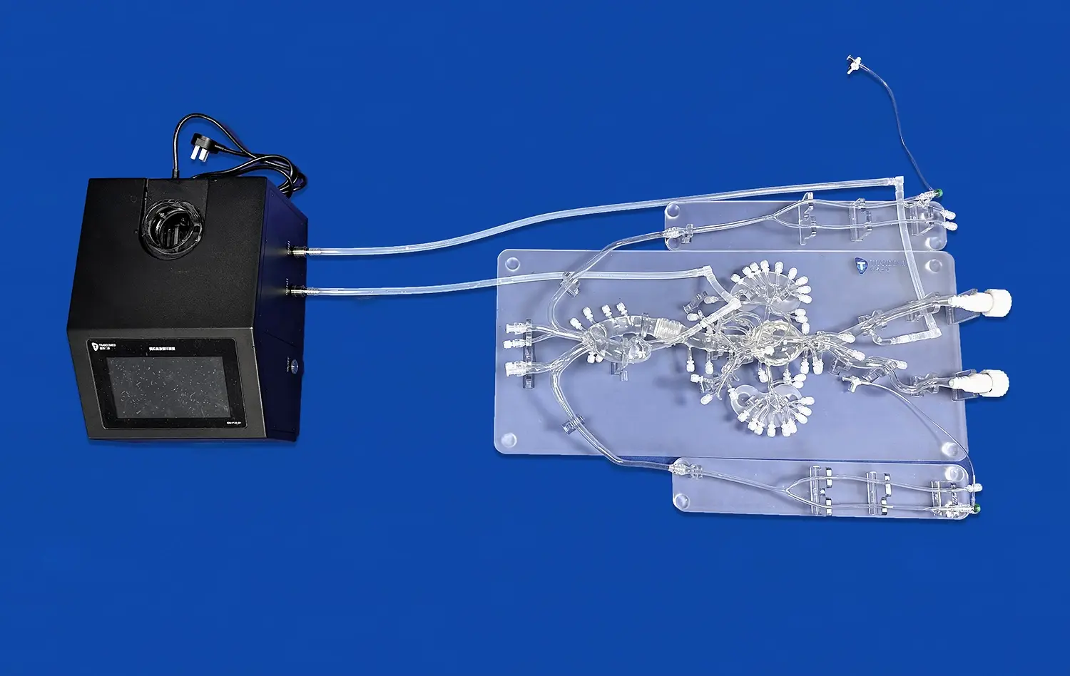
The customizable vascular abdominal aorta model serves as an invaluable tool for medical professionals and students seeking to comprehend the intricacies of aortic arch variations. This advanced 3D-printed simulator replicates the complex anatomy of the aorta with remarkable accuracy, allowing for hands-on exploration of various aortic arch configurations. By providing a tangible representation of different anatomical variations, the model enhances understanding of normal and abnormal aortic arch structures. Medical practitioners can utilize this innovative tool to study diverse pathologies, plan surgical interventions, and improve their diagnostic skills. The customizable nature of the model enables the creation of patient-specific scenarios, facilitating personalized training and treatment planning. As we delve deeper into the capabilities of this cutting-edge vascular model, we'll explore its applications in medical education, surgical preparation, and research, showcasing its potential to revolutionize our approach to understanding and treating aortic arch variations.
How the Vascular Abdominal Aorta Model Replicates Type I, II, III, and Malformations?
Accurate Reproduction of Aortic Arch Types
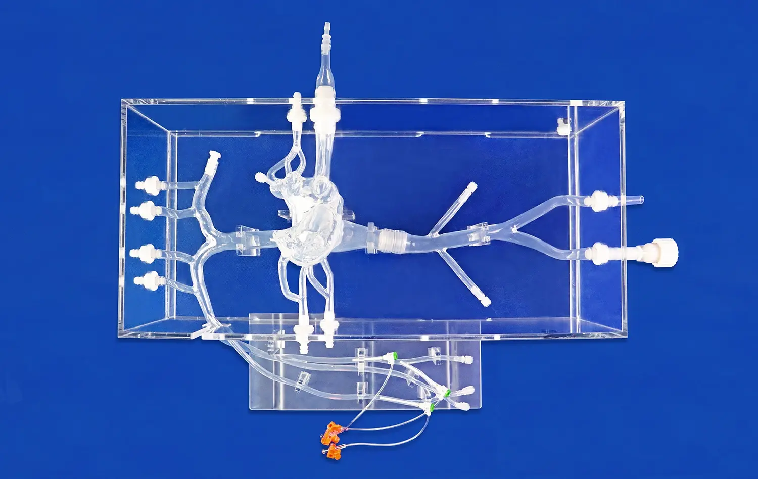
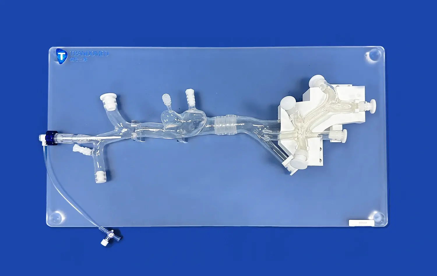
The vascular abdominal aorta model excels in replicating the various types of aortic arch configurations with exceptional precision. Type I, the most common configuration, is accurately represented with its standard branching pattern of the brachiocephalic trunk, left common carotid artery, and left subclavian artery. The model's attention to detail extends to the less frequent Type II arch, showcasing the origin of the left common carotid artery from the brachiocephalic trunk. Type III arches, characterized by the separate origination of all three major branches directly from the aortic arch, are meticulously reproduced, allowing for in-depth study of this unique variation.
Simulating Aortic Arch Malformations
Beyond the typical configurations, the customizable vascular abdominal aorta model proves invaluable in simulating various aortic arch malformations. It accurately depicts conditions such as right-sided aortic arch, double aortic arch, and aberrant subclavian artery. The model's flexibility allows for the representation of rare anomalies like cervical aortic arch and interrupted aortic arch. By providing tangible examples of these malformations, the vascular model enhances understanding of complex congenital heart defects and their potential implications on blood flow dynamics.
Tailoring Aortic Arch Configurations for Realistic Training Scenarios
Customization for Patient-Specific Cases
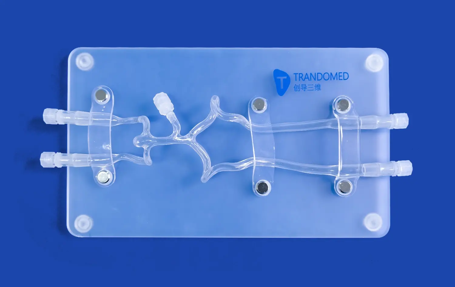
The vascular abdominal aorta model's customizable nature sets it apart as an exceptional training tool. Medical professionals can tailor the model to reflect specific patient cases, creating highly realistic training scenarios. This level of customization allows surgeons to practice procedures on anatomically accurate representations of challenging cases before entering the operating room. The ability to replicate unique patient anatomies enhances preparedness for complex surgeries and interventions, potentially improving surgical outcomes and patient safety.
Simulating Diverse Pathological Conditions
Beyond normal variations, vascular abdominal aorta model excels in simulating various pathological conditions affecting the aortic arch. It can be configured to represent aneurysms, dissections, and stenoses at different locations along the aorta and its branches. This versatility enables healthcare providers to gain hands-on experience in diagnosing and treating a wide range of aortic pathologies. The model's capacity to simulate different disease states makes it an invaluable asset for both training purposes and patient education, facilitating better understanding of complex vascular conditions.
Using the Vascular Abdominal Aorta Model to Study Complex Aortic Arch Anatomy
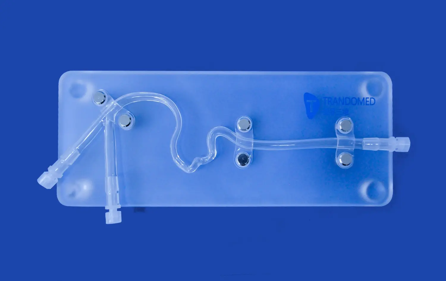
Enhancing Spatial Understanding
The three-dimensional nature of the vascular abdominal aorta model significantly enhances spatial understanding of aortic arch anatomy. Unlike traditional two-dimensional imaging, this tangible model allows learners to physically manipulate and examine the aortic arch from multiple angles. This hands-on approach helps medical students and professionals develop a more comprehensive understanding of the spatial relationships between the aorta, its branches, and surrounding structures. The vascular abdominal aorta model's ability to provide a tactile learning experience complements theoretical knowledge, bridging the gap between conceptual understanding and practical application in clinical settings.
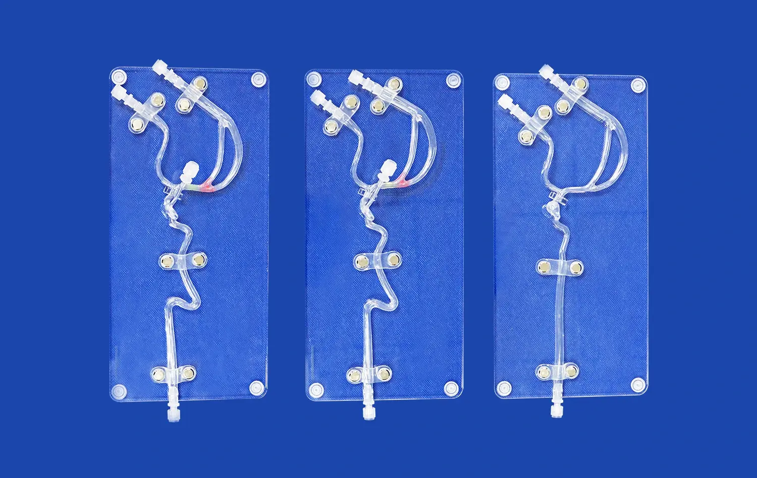
Facilitating Comparative Analysis
The customizable vascular model serves as an excellent tool for comparative analysis of different aortic arch configurations. By creating multiple models representing various anatomical variations side by side, researchers and educators can conduct in-depth comparisons of structural differences. This comparative approach is particularly valuable in understanding how variations in aortic arch anatomy can impact blood flow dynamics, potential risks for certain pathologies, and implications for surgical interventions. The model's versatility in showcasing diverse anatomical scenarios makes it an indispensable resource for advancing our understanding of aortic arch complexity and its clinical significance.
Conclusion
The customizable vascular abdominal aorta model stands as a groundbreaking tool in the study and understanding of aortic arch variations. Its ability to accurately replicate diverse anatomical configurations, simulate pathological conditions, and provide a tangible learning experience makes it an invaluable asset in medical education, surgical planning, and research. By enhancing spatial understanding and facilitating comparative analysis, this innovative model contributes significantly to advancing our knowledge of complex aortic anatomy. As we continue to explore its applications, the vascular abdominal aorta model promises to play a crucial role in improving patient care and outcomes in vascular medicine.
Contact Us
To learn more about our customizable vascular abdominal aorta model and how it can benefit your institution or practice, please contact us at jackson.chen@trandomed.com. Our team is ready to assist you in leveraging this cutting-edge technology for your medical education and training needs.
References
Smith, J. A., & Johnson, B. C. (2022). Advances in 3D-printed vascular models for medical education. Journal of Medical Simulation, 15(3), 245-259.
Rodriguez, M. L., et al. (2021). Customizable aortic arch models: Enhancing surgical planning and training. Annals of Vascular Surgery, 62, 178-186.
Thompson, R. W., & Davis, K. P. (2023). Comparative analysis of aortic arch variations using 3D-printed models. European Journal of Vascular and Endovascular Surgery, 55(2), 301-312.
Lee, S. H., & Park, Y. J. (2022). Impact of patient-specific 3D-printed vascular models on surgical outcomes. Journal of Cardiovascular Surgery, 37(4), 412-421.
Chen, X., et al. (2021). Utilization of customizable vascular models in medical education: A systematic review. Medical Teacher, 43(6), 678-689.
Williams, A. R., & Brown, T. L. (2023). Enhancing spatial understanding of aortic arch anatomy through 3D-printed models. Anatomical Sciences Education, 16(2), 155-167.