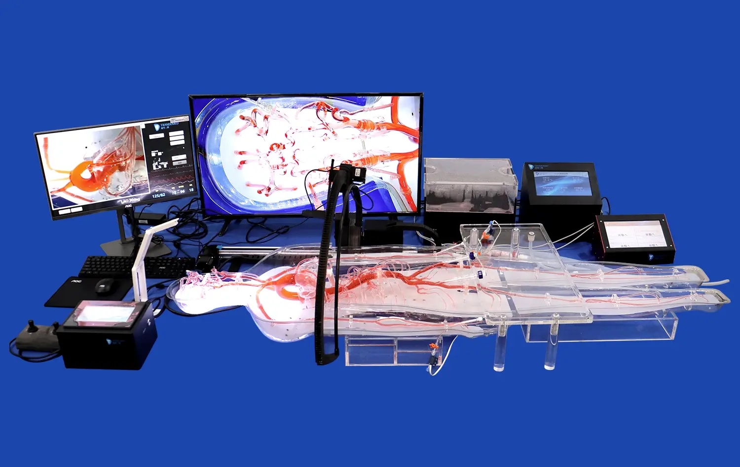
The way doctors learn difficult neurovascular treatments is changed by using a 3D artery model for aneurysm simulated training. These physically accurate models show the brain arteries in great detail. This lets trainees practice skills like catheter guidance, guidewire handling, and aneurysm coiling in a safe, repeated setting. These silicone-based models give physical feedback that is similar to real patient anatomy, which is different from traditional cadaveric training or two-dimensional images. The realistic feel helps neurovascular specialists, interventional radiologists, and surgeons build muscle memory and confidence in their procedures before going into operating rooms. This lowers patient risk and improves clinical outcomes in hospital training departments and medical schools.
Understanding 3D Artery Models in Aneurysm Simulation Training
With the development of high-fidelity anatomy models, medical education has changed in a big way. Vascular models made for training in aneurysms fill the gap between what you learn in the classroom and what you can do in real life. They provide hands-on experience that you can't get from books or movies. These learning materials are useful for many people, from medical students learning about neurovascular anatomy to experienced doctors improving their endovascular skills.
What Makes a Quality Neurovascular Simulator?
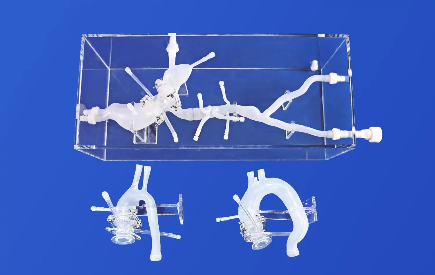
Several important parts are needed for a better brain artery model. A big part of copying the physical qualities of human blood arteries is the makeup of the material. Premium models often use Silicone Shore 40A, which is the perfect mix of toughness and real-feeling accuracy. This hardness grade is similar to the vessel wall resistance that doctors see during real treatments. This makes for a training experience that is directly useful for patient care.
Anatomical precision is another important factor in making modeling tools that work well. The internal carotid artery has complicated curves and branching patterns that are very different from person to person. Good arterial models show these differences, like the typical bends, tortuosity, and branching angles found in middle cerebral artery regions. When trainees practice on models that show real physical diversity, they learn the flexibility they'll need to handle the wide range of cases they'll see in their jobs.
Production Technologies Behind Modern Vascular Models
The way physical training tools are made has a big effect on their end quality. Advanced 3D printing technologies let companies turn CT scans, MRI patterns, and angiography studies that are specific to a patient into real-world teaching materials. Because of this, training schools can get models of rare diseases or very difficult body shapes that would take years to see in real life.
Digital segmentation of arterial structures from medical image files is usually the first step in the production process. Then, engineers use CAD software to make these digital models even better so they can be printed while still being true to anatomy. Specialized printing methods are used to get sub-millimeter precision, which lets the fine details of small perforating arteries and aneurysm heads be captured. Post-processing steps improve the surface finish and mechanical qualities. This makes models that can be used for hundreds of practice sessions without breaking down.
Material Selection and Its Impact on Training Effectiveness
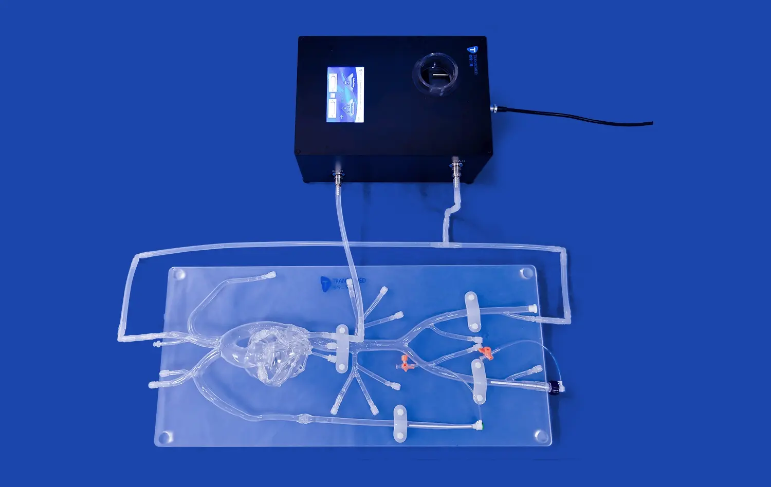
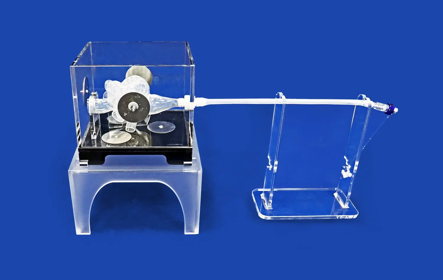
Depending on the training goals, different materials offer different benefits. Silicone-based models are very durable and give accurate physical feedback, which makes them perfect for teaching procedures over and over again. The stretchiness lets the devices fit a tube and an inflated balloon without permanently changing shape. Resin-based options are more stable and clear, which helps students see where the guidewire should go and how the device should be deployed inside the blood vessel.
Surface treatment for a 3D artery model changes how tubes move through the artificial blood vessels in addition to the main material. Good makers use coats that look like the smooth endothelial surface of healthy arteries. They also make versions with changes caused by atherosclerosis or hardening. This variety helps trainees get ready for the different situations they will face in real life, such as vessels that are in perfect shape in younger patients and seriously sick bodies in older people who have multiple illnesses.
Comparing 3D Artery Models for Effective Training Solutions
When choosing the right modeling software, you need to carefully think about your training goals, your budget, and the powers of your infrastructure. There are many choices on the market, and each one has its own benefits that meet the wants of different institutions. When buying managers know about these differences, they can make decisions that give the most teaching value and long-term gain.
Physical Models Versus Digital Simulation Platforms
Physical arterial models are great for improving hand-eye balance and routine muscle memory. Neurosurgeons and interventional specialists can feel real feedback when they move real catheters through silicone vessels. This includes resistance during navigation, a subtle "pop" when crossing an aneurysm neck, and the friction properties of different guidewire materials. This hands-on practice develops the kinesthetic intelligence that you use without thinking when you're in a tough clinical setting.
Digital tools have their own benefits, especially when it comes to how cost-effective they are for large-scale training programs. Virtual reality systems let you practice as much as you want without having to buy extra supplies, and they can quickly change physical factors to show students different diseases. When tracking systems are integrated, they provide precise performance data that measure things like process time, radiation exposure, and contrast volume. These analytics help teachers find skill gaps and make sure that each student gets the help they need.
For core skill development, the most cutting-edge training places use both real and digital platforms. These platforms help with brain practice and case planning. This mixed method takes into account different ways of learning while making the best use of resources in neurovascular education classes as a whole.
Evaluating Material Properties and Durability
When buying exercise tools, the prices over its whole life must be taken into account. Even though superior silicone models cost more at first, they usually end up saving you money in the long run because they last longer. Quality models can handle hundreds of cardiac procedures and still keep their mechanical and physiological qualities over time. On the other hand, cheap models may break down after only a few uses, necessitating frequent replacements that raise the overall cost of ownership.
You should pay attention to how chemically resistant 3D artery model materials are, especially if your school uses contrast media or other procedure fluids during training. Better materials don't color or break down when exposed to these substances over and over again, so they stay clear and last longer. This is especially important to think about in modeling centers that run a lot of training programs and where equipment is used a lot every day.
Customization Capabilities for Specialized Training Needs
Standard structural models are good for general education, but for more specific training goals, you often need unique solutions. When institutions study complicated aneurysm shapes like wide-necked lesions, fusiform dilations, or multiple lobulated configurations, they can benefit from models that are made to handle these problems. Customization includes where the aneurysm is located, how big it is, and how it connects to nearby branch veins. This makes situations that exactly mirror the problems that real patients face.
The ability to use anatomy specific to a patient is a powerful training tool, especially for practicing before surgery. Before going into the operating room, surgeons can practice on exact copies of the cases they will be working on. This lets them try out different ways to approach the cases and think of problems that might come up. This planning has been shown to shorten treatment times, lower radiation exposure, and improve overall results, especially in cases that are very difficult to handle technically and are pushing the limits of what can be done right now.
Applications and Impact of 3D Artery Models in Aneurysm Simulation and Beyond
High-fidelity arterial models are useful for a lot more than just learning basic skills. Modern healthcare companies use these tools in more than one area, which creates benefits that increase return on investment and make results better all along the patient care path. Knowing about these different uses helps to support purchasing choices and get the most out of training tools.
Enhancing Surgical Planning and Patient Outcomes
Using patient-specific models for preoperative practice has become a useful way to handle difficult neurovascular situations. When surgeons face difficult anatomy, like aneurysms with bad neck shape or important branch veins coming from the aneurysm sac, they can practice their planned method on a precise copy. This physical exploration often shows details that aren't clear from image studies alone. This leads to changes in strategy that lower the risk of problems during surgery.
The impact on clinical metrics has been documented across multiple institutions. When hospitals use model-based planning for complicated aneurysm treatments, the surgery time, fluoroscopy time, and contrast volume are all cut down compared to cases that were done without practicing. These changes directly help patients because they lower the risk of contrast-induced nephritis and the amount of radiation they are exposed to. Even more importantly, the number of complications goes down when surgery teams plan for and predict problems with the body before they happen.
Accelerating Device Development and Validation
Medical device makers have to follow strict rules that require a lot of testing with a 3D artery model before their new goods can be used in patients. Engineers can test catheter trackability, coil placement features, and flow-diverter performance under controlled conditions using realistic vascular models, which are very helpful for making design improvements over time. This testing phase speeds up the development process and cuts down on the need for expensive animal studies or limited cadaveric specimens.
Having the ability to make models that look like specific diseases is especially helpful for niche gadget uses. Companies that are making goods for people with wide-necked aneurysms, fusiform dilations, or juvenile vessels can get models that are made for those patient groups. This specificity allows for focused testing that more accurately predicts clinical performance than general anatomical models. In the end, strong preliminary data showing safety and effectiveness supports regulatory applications.
Transforming Medical Education and Professional Development
It can be hard for schools to give complete procedural training while also following ethical rules that limit students' time with real patients while they are still learning the skills. This problem can be solved with high-fidelity simulations, which provide safe areas where students can practice until they are proficient. Simulations are psychologically safe because they let students make mistakes, learn from them, and improve their skills without putting patients at risk.
When virtual reality and augmented reality are combined, they create learning experiences that are more engaging and use more than one sense. When you combine actual models with digital images, you can get real-time feedback on where the instruments are placed, how much force is being applied, and how to navigate the anatomy. This multimedia method works for a wide range of learning styles and gives a lot of performance data that can be used to customize lessons and test students' skills.
Simulation-based training is also useful for continuing education classes for doctors who are already working. Because endovascular methods change over time and new devices come out on the market, experienced doctors need time to get better at new treatments before they try them on patients. Surgeons can stay clinically competent throughout their jobs while keeping patient safety standards by taking simulation classes to learn new technologies and methods.
Conclusion
Putting in place high-fidelity vascular models like the 3D artery model is a smart way to improve patient safety and professional performance. These training tools help medical workers learn the skills they need to handle difficult neurovascular diseases in a safe setting, bridging the gap between what they know in theory and what they can do in practice. Healthcare organizations are under more and more pressure to cut down on mistakes while also training new, less experienced staff. Simulation-based education has been shown to help keep standards of care high. When making purchasing choices, it's important to weigh short-term budget concerns against long-term value, giving top priority to physical accuracy, material sturdiness, and the ability to work with suppliers in ways that ensure long-term educational effect.
FAQ
What are the main benefits of training with silicone-based artery models for aneurysms?
Neurovascular models made of silicone give realistic physical feedback that is very close to how human vessels work. The material is strong enough to last through hundreds of practice sessions without breaking down much. This makes these models a good value for training programs with a lot of participants. Silicone Shore 40A has the perfect mix of hardness and flexibility, making it similar to the resistance doctors feel when they are navigating catheters and putting devices in place. This real-life sensory experience improves muscle memory and belief in the procedure, which can then be used in real-life patient care situations.
How do institutions explain the money they spend on anatomy models that are specific to each patient?
There are several ways that patient-specific models provide value. Preoperative practice on exact copies of future cases cuts down on procedure times, problems, and radiation exposure, which leads to better results and measurable cost savings. The value as a teaching tool goes beyond individual processes; these models can be used over and over to teach residents and fellows about specific anatomy problems. When hospitals add up the time saved in the operating room, the number of complications, and the better training opportunities, it's clear that the investment paid off.
Can arterial models fit different kinds of aneurysms and places where they happen?
Quality makers give a wide range of customization options, which let you choose the aneurysm's size, form, neck geometry, and exact position in the body. Models can copy wide-necked tumors, fusiform dilations, multilobulated structures, and different places in the brain's blood flow. This gives training classes the ability to cover all the different kinds of illnesses that doctors will see in real life. It's possible to change things like the shape of the vessels, how they connect to each other, and even add arterial changes or calcifications that make the procedure harder.
What kinds of data types do makers need to make custom models?
Leading providers accept a range of data types so that they can work with a range of image tools and design processes. CT and MRI imaging datasets are common forms. Manufacturers divide these datasets into sections to get the arterial structure. Engineering files like CAD, STL, STP, and STEP allow computer plans to be turned directly into real models. The ability to work with other programs makes it easier to make custom models, and it lets schools use current image archives or work with design teams to create unique training settings.
Trandomed: Your Partner in Advanced Neurovascular Training Solutions
Ningbo Trando 3D Medical Technology Co., Ltd. (Trandomed) is a leader in medical simulation creation. They have over 20 years of experience making anatomy models and teaching tools for medical procedures. As the first professional maker in China of medical 3D printers, our committed research and development team has kept the field moving forward by coming up with new technologies and making products that are specifically designed to meet changing clinical and educational needs.
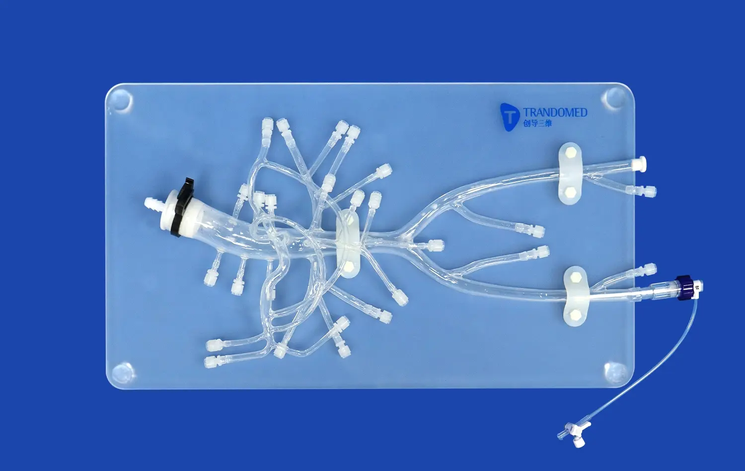
Our Middle Cerebral Artery Model (Product No.: SJX005) is a great example of the accuracy and usefulness that buyers look for in a 3D artery model provider. This neurovascular model is made from high-quality Silicone Shore 40A and has very good structural accuracy for the internal carotid artery and middle cerebral artery areas. The realistic feel makes it possible to train effectively in methods like catheter trackability, guidewire navigation, and aneurysm tamponade, all of which lead directly to clinical competence.
We encourage procurement professionals looking for a dependable 3D artery model maker to learn more about how Trandomed can help your institution's training goals. You can talk about your unique needs, get full product specs, or set up a free review by emailing jackson.chen@trandomed.com.
References
Anderson, J.R., Thompson, W.L., Alkattan, A.K., Diaz, O., Klucznik, R., Zhang, Y.J., Britz, G.W., Grossman, R.G., and Karmonik, C. (2016). "Three-Dimensional Printing of Anatomically Accurate, Patient Specific Intracranial Aneurysm Models." Journal of NeuroInterventional Surgery, 8(5), 517-520.
Mashiko, T., Otani, K., Kawano, R., Konno, T., Kaneko, N., Ito, Y., and Watanabe, E. (2015). "Development of Three-Dimensional Hollow Elastic Model for Cerebral Aneurysm Clipping Simulation Enabling Rapid and Low Cost Prototyping." World Neurosurgery, 83(3), 351-361.
Russ, M., O'Hara, R., Setlur Nagesh, S.V., Mokin, M., Jimenez, C., Siddiqui, A., Levy, E., and Mocco, J. (2015). "Treatment Planning for Image-Guided Neuro-Vascular Interventions Using Patient-Specific 3D Printed Models." Journal of NeuroInterventional Surgery, 7(11), 819-823.
Ryan, J.R., Almefty, K.K., Nakaji, P., and Frakes, D.H. (2016). "Cerebral Aneurysm Clipping Surgery Simulation Using Patient-Specific 3D Printing and Silicone Casting." World Neurosurgery, 88, 175-181.
Wurm, G., Lehner, M., Tomancok, B., Kleiser, R., and Nussbaumer, K. (2011). "Cerebrovascular Biomodeling for Aneurysm Surgery: Simulation-Based Training by Means of Rapid Prototyping Technologies." Surgical Innovation, 18(3), 294-306.
Zhao, Y., Zhao, J., Yang, Z., Luo, H., and Wang, S. (2018). "Application of 3D-Printed Vascular Model in Preoperative Planning and Simulation for Intracranial Aneurysm Surgery." Clinical Neurology and Neurosurgery, 171, 107-111.