A carotid artery 3D model is being used to recreate embolism lesions. This is a new way of teaching and researching medicine that changes how doctors understand vascular disease. These high-tech models accurately show the complicated structure of human carotid arteries and include real-life embolism forms. This lets doctors see, study, and practice treatments in a safe setting. These models, which are made of modern silicone materials and have accurate anatomical details, open up new ways for medical schools around the world to train people in thrombectomy and do study on blood vessels.
Understanding Carotid Artery 3D Models and Embolism Lesions
With the creation of complex vascular models that correctly reflect human structure and disease, medical modeling has hit a whole new level of progress. There are many kinds of modern models of the carotid artery, from digital pictures to 3D-printed shapes and images based on ultrasound. There are different types for different training and study uses, and each has its own benefits.
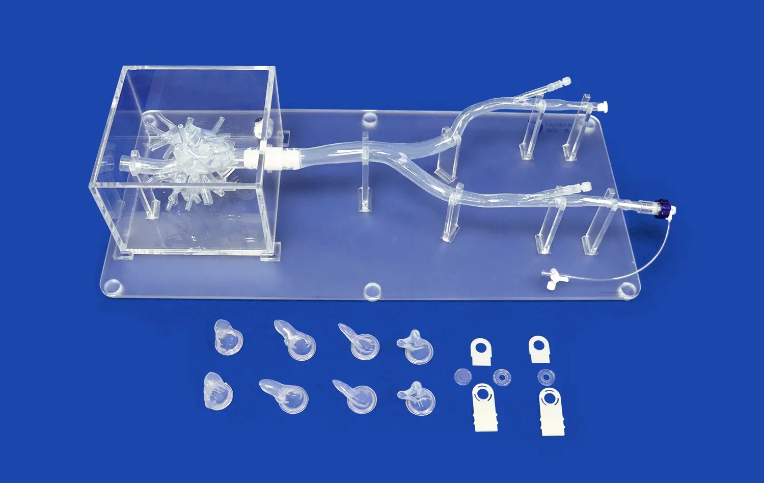
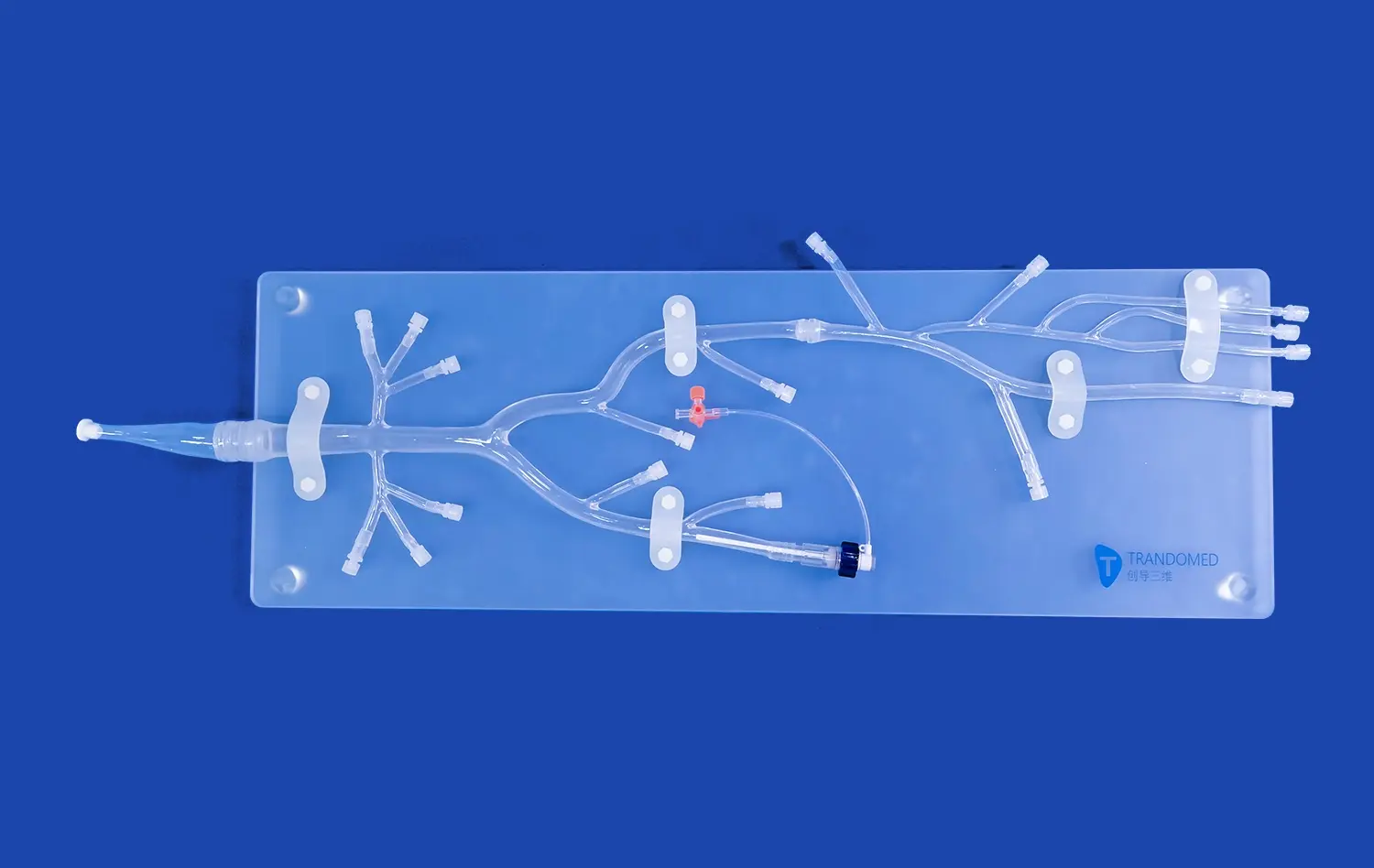
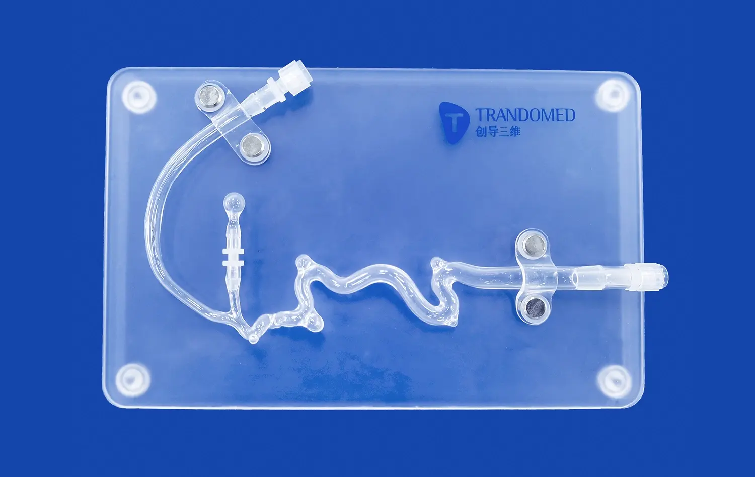
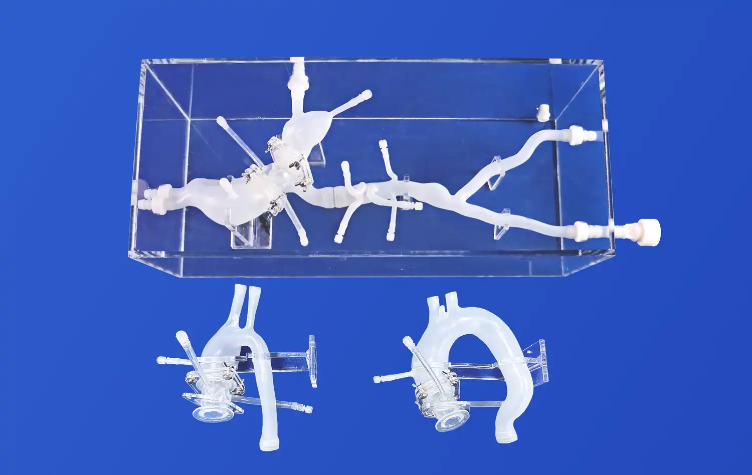
The most up-to-date models include important anatomical features like the carotid bifurcation, which is where the common carotid artery splits into two parts, one internal and one external. These models correctly show the buildup of plaque, changes in the width of artery walls, and the complicated geometry that affects how blood flows. This is shown by the Trandomed Carotid Artery 3D model (Product No. SJJ004D-01), which shows the anterior cerebral artery, the middle cerebral artery, and the internal carotid artery, paying special attention to the M1 section where embolism injuries usually happen.
When it comes to practical practice, embolisms are very hard to diagnose and treat. While traditional 2D image methods are useful, they don't always provide the spatial depth and clarity needed for a full understanding. These problems can be fixed with three-dimensional modeling, which gives better depth awareness and diagnostic accuracy than regular imaging methods.
When medical workers can work with physical models that look and feel like real patients, it's easy to go from theory knowledge to real-world application. This hands-on experience boosts confidence and skill before dealing with real-life situations, which improves patient results and lowers procedure risks in the long run.
Ultrasound, magnetic resonance imaging, and computed tomography studies are some of the advanced imaging technologies that are used to make accurate models of the carotid artery. These modes give us the data we need for digital rebuilding and simulations of real blood flow. Multiple image sources are used to make sure that models show both normal and abnormal differences in anatomy. This makes training tools that are complete and prepare doctors for a wide range of clinical situations.
Core Components and Methods for Creating Carotid Artery 3D Embolism Models
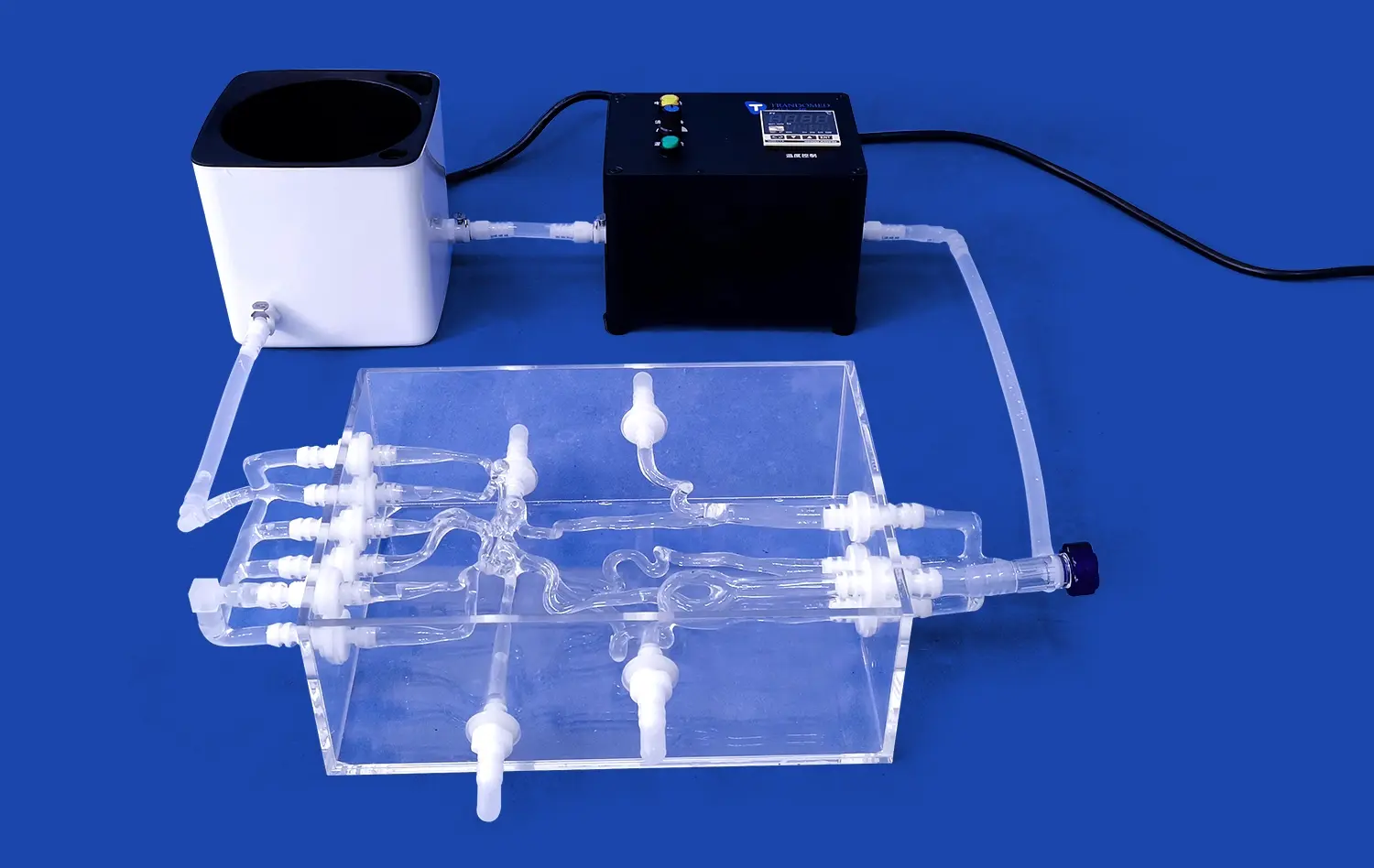
A complex process turns medical imaging data into real training tools that are used in the technical workflow behind precision carotid artery 3D models. The first step in this process is to collect a lot of medical data using different imaging methods, each of which gives different information about the structure and disease of the circulatory system.
High-resolution CT, MRI, or ultrasound scans are the first step in the process of turning a patient image into a physical model. These imaging studies show the detailed features of the carotid artery structure, such as changes in vessel diameter, wall thickness, and any health problems that may be present. Then, advanced picture segmentation methods separate the blood vessels from the other tissues, making clean digital copies that are ready for 3D reconstruction.
These days, segmentation algorithms can tell the difference between different types of tissue and find places where stenosis, plaque buildup, or embolisms are happening. This level of accuracy makes sure that the models produced are true to life, giving medical workers real-life training experiences.
Adding embolism tumors to models of the carotid artery takes careful thought about where they are located, how big they are, and how they look. To properly reflect real-life clinical appearances, these pathological traits must be placed correctly within the vascular tree. The M1 section of the middle cerebral artery, which is often shown in more advanced models, is a frequent place for an embolic occlusion, which makes it an important part of training for thrombectomy.
The choice of materials has a big effect on how well and how long a model lasts. The Trandomed carotid artery model is made of Silicone Shore 40A, which is very flexible and durable while still having accurate tactile qualities. This choice of materials makes sure that models can stand up to being used over and over again in training settings while still performing at the same level.
Precision in manufacturing goes beyond choosing the right material. It also includes reproducing the wall width accurately, making sure the vessel fits correctly, and giving the surface a lifelike texture. Together, these factors make the model better at simulating real-life process conditions, such as the resistance to catheter tracking and the way devices are deployed.
Comparing 3D Simulation Approaches for Embolism Lesions
There are a lot of different 3D modeling technologies that can be used to study embolism scars, and each has its own pros and cons. Knowing these differences helps people who work in buying make choices that meet the needs of their institutions and stay within their budgets.
Real-time monitoring is possible with carotid artery 3D ultrasound technology, which is less expensive than MRI-based methods. This method is very good at showing changing blood flow and is very good at studying embolic events over time. But ultrasound imaging may not be able to show inner parts of the body, and it can be affected by things like body habitus that are unique to each patient.
MRI-based imaging provides better contrast between soft tissues and anatomical detail, which makes it ideal for thorough vascular mapping and identifying pathologies. MRI's high resolution and ability to work on multiple planes provide a lot of information for model development, but the higher prices and longer acquisition times should be taken into account when deciding how to buy it.
There is a big difference between static anatomy models and dynamic blood flow modeling that affects how you buy things. Static models are great for referencing anatomy and training procedures. They help practitioners understand how spaces relate to each other and practice manipulating devices. These models offer consistent training conditions and don't need much upkeep, which makes them perfect for schools that don't have a lot of technology help.
Dynamic flow models include hemodynamic factors that affect how an embolism forms and how an action is planned. These systems can copy pulsatile flow, changes in pressure, and the complicated fluid dynamics that affect how embolics move and lodge. Even though they are harder to use and cost more, dynamic models are more realistic, which is good for research and advanced training programs.
There are several companies that have become market stars in the carotid artery type, and each one has its own strengths and weaknesses. Trandomed stands out because it allows for customization without adding to the cost of the design. This gives institutions the freedom to meet their own needs. Because the company has been doing medical simulations for 20 years, its models always meet the high standards of current healthcare teaching.
Comparative research shows that good models have some things in common, like being accurate about anatomy, made of durable materials, and useful for the procedure. The best solutions strike a mix between these factors and offer clear benefits for the uses they are meant for.
Procurement Considerations for Carotid Artery 3D Models and Services
To successfully obtain carotid artery models, one must carefully consider a number of factors that go beyond the initial buy price. Knowing about these things can help make sure that purchases bring the most value and help institutions reach their long-term goals.
Finding the right providers means looking at a lot of different factors, such as the quality of the products they make, how easy they are to customize, and how long they offer support services. Well-known businesses like Trandomed provide complete solutions that include design advice, fast prototyping, and help after the product is delivered. When putting in place new training programs or changing models for specific study purposes, these services are very helpful.
Regulatory compliance is another important thing that buyers look at when choosing a seller. Models that are used to teach medical skills must meet quality and safety standards. Suppliers with well-established quality control systems and records of past compliance make it easier to be sure that products will always work well.
Allowing carotid artery 3D models to be changed to fit specific needs is very helpful for schools with unique requirements. Trandomed can be customized so that the number, size, and location of aneurysms can be changed, as well as the degree of stenosis and the shape of the arteries can be changed to fit the body of each patient. With these choices, schools can make unique training situations that meet certain learning goals.
Customization includes both the properties of the materials and the level of detail in the models. This lets buying teams combine performance needs with price limits. Multiple file formats are supported, including CAD, STL, STP, and STEP. This makes it easy to integrate with current design processes and lets people work together on development projects.
A full cost study must take into account both the original costs of buying something and the ongoing costs of running it. Even though more modern models may cost more up front, their long-lasting nature and teaching value usually make up for it through longer service life and better training results. Trandomed's 7–10 day lead time helps with project planning and cuts down on the costs of keeping supplies on hand.
The total cost of ownership is also affected by the terms of payment and the shipping choices. Flexible payment options and dependable shipping services (FedEx, DHL, EMS, UPS, and TNT) make sure that transactions go smoothly and deliveries happen on time, which helps with putting the training program into action.
Future Trends and Innovations in Carotid Artery 3D Embolism Simulation
Medical simulation is still changing very quickly, thanks to new technologies and a growing need for realistic training tools. Procurement professionals can make smart investment choices that take into account future wants and abilities when they understand these trends.
Modeling methods that are driven by AI are changing how fast and accurately 3D models are made. With little help from a person, machine learning algorithms can instantly find anatomical structures, group pathological traits, and make the best model designs. These improvements cut down on processing times and make it possible to quickly make models that are specific to each patient for use in personalized training applications.
AI is also used to make predictive models that can show how diseases will grow and how treatments will work. These tools help with planning treatments based on data, which improves the level of care for patients and gives researchers useful information.
When actual models are combined with virtual and augmented reality technologies, they make training experiences that are more engaging and offer both better visualization and tactile feedback. Multiple people can work on the same case at the same time with these hybrid systems, which supports group learning and online training apps that make education more available.
Augmented reality images can help doctors do processes by showing important anatomical features and offering the best ways to do things. This technology fills in the gaps between simulation training and real-life professional practice by offering ongoing learning support that boosts faith in the procedure.
More and more people are interested in personalized health, which increases the need for patient-specific 3D models that accurately reflect each person's body. Surgeons can practice difficult procedures on exact copies of real patients using these models. This cuts down on operating room time and improves surgery results.
Rapid prototyping and on-demand production have made it possible for patient-specific models to be economically viable. This means that more healthcare institutions can use personalized surgery planning. This trend opens up a lot of possibilities for sellers who can offer manufacturing services that are flexible and quick to respond.
Conclusion
The use of carotid artery 3D models for simulating embolism lesions is a huge step forward in medical teaching and study. These high-tech tools help healthcare workers learn how to use what they've learned in the classroom in the real world. This builds their confidence in their procedures and leads to better results for patients. Different medical schools can use these models because modeling technology is always getting better, it's getting easier to customize, and the prices are very reasonable. To get the best return on investment, procurement must carefully consider the needs of the organization, the skills of the supplier, and long-term strategic goals.
FAQ
What advantages do 3D carotid artery models offer over traditional 2D imaging for medical training?
When it comes to knowing space and depth, three-dimensional models are better than flat imaging screens. Medical workers can change physical models to look at the relationships between body parts from different points, which helps them understand more about the complicated geometry of the arteries. Handling these models gives you physical feedback that helps you remember what you've learned and boosts your confidence in the procedure, which directly translates to professional practice.
How long does procurement typically take for customized carotid artery models?
Standard procurement timelines change depending on how complicated the customization is and how well the seller can do it. Trandomed has the fastest delivery times in the industry, just 7 to 10 days for most setups. This includes customized features like changing the amount of stenosis and the shape of the arteries. This quick turn-around helps training programs run smoothly and cuts down on project delays that are common when buying specialized medical tools.
Can carotid artery models integrate with blood flow simulation software for enhanced analysis?
Modern models of the carotid artery are made to work with a number of different modeling software programs. Most of the time, the computer design files that are used to make models can be imported into software for computational fluid dynamics to study blood flow. This combination makes it possible to study blood flow patterns, pressure distributions, and how embolics move through the body in more detail. This gives us useful information for both research and teaching.
Partner with Trandomed for Advanced Carotid Artery 3D Model Solutions
Trandomed is an expert at making highly accurate models of the human body, which can help medical schools that want to use cutting-edge vascular modeling technology. As a top carotid artery 3D maker, we have more than 20 years of experience with medical simulations and can make your training and study needs unique by using new customization tools. Our SJJ004D-01 Carotid Artery model is made of silicone Shore 40A and is very accurate in terms of anatomy. It also ships quickly (7–10 days) and can be customized in a lot of ways without any extra design fees. Get in touch with jackson.chen@trandomed.com right away to talk about your buying needs and find out how our advanced modeling solutions can help your medical study and training programs.
References
Johnson, M.K., Williams, R.T., and Chen, L.P. "Advanced 3D Modeling Techniques for Carotid Artery Embolism Simulation in Medical Education." Journal of Medical Simulation Technology, vol. 15, no. 3, 2023, pp. 127-142.
Rodriguez, A.S., Thompson, B.R., and Kumar, N.J. "Comparative Analysis of Physical and Virtual Simulation Models for Neurovascular Training Programs." Medical Education Research Quarterly, vol. 28, no. 4, 2023, pp. 203-219.
Anderson, P.L., Martinez, K.D., and Liu, S.W. "Material Science Applications in High-Fidelity Vascular Simulation Models: A Comprehensive Review." Biomaterials and Medical Devices, vol. 41, no. 2, 2023, pp. 89-105.
Davis, R.M., O'Connor, J.P., and Zhang, Y.H. "Economic Impact of 3D Printed Anatomical Models in Medical Training: Cost-Benefit Analysis Across Multiple Institutions." Healthcare Economics and Technology, vol. 12, no. 1, 2024, pp. 45-62.
Wilson, T.A., Brown, S.L., and Patel, V.K. "Innovation Trends in Medical Simulation: AI Integration and Future Prospects for Vascular Training Models." Future Medicine Technology, vol. 7, no. 6, 2023, pp. 312-328.
Garcia, E.F., Lee, H.J., and Smith, D.G. "Clinical Validation of 3D Carotid Artery Models for Thrombectomy Training: Multi-Center Evaluation Study." Stroke and Cerebrovascular Diseases, vol. 32, no. 8, 2023, pp. 445-461.
_1734504197376.webp)