Using a Circle of Willis Brain Model for Neuro-Interventional Education
2026-03-06 09:00:04
The circle of willis brain model is an important learning tool for doctors who are trained in neurointerventional procedures because it accurately shows the anatomy of the cerebral vasculature. The complex network of arteries that bring blood to the brain is shown correctly in these high-tech models. With this, students can learn by doing, which isn't possible with regular books. High-fidelity brain models are used by medical schools all over the world to improve their training programs, make surgeries more successful, and move neurovascular medicine studies forward. Because these models are so true to life, they help professionals and students learn about important blood vessel pathways, practice interventional treatments, and get better at taking care of neurovascular conditions that are hard to manage.
Understanding the Circle of Willis Brain Model
Anatomical Structure and Function
The Circle of Willis is a triangle-shaped network of arteries at the base of the brain that connects the front and back cerebral circulations. These arteries can talk to each other. This blood vessel band is made up of the internal carotid arteries, the anterior cerebral arteries, the anterior communicating artery, the posterior cerebral arteries, and the posterior communicating arteries. The structure's unique shape makes two sets of blood supply routes. This way, even if some blood vessels get stopped or narrow, the brain will still get blood.
Models of the modern brain correctly show this complicated anatomy by using vessel diameters, branching patterns, and spatial relationships that are similar to how the human body works. You can teach a lot more than just basic anatomy with these models. They show people how blood moves, how body parts work together to help, and how differences in physique can impact health. Students can learn about how different diseases can affect brain blood flow and the different ways that surgery can be used for different types of treatments.
Educational Applications in Neurovascular Training
These models can be used to help medical students learn in a number of settings that help them understand better and get better at what they're doing. These tools are used in training classes to show how the vascular system should look, to make it look like it is sick, and to practice interventional methods in a safe environment. Students' sense of space gets better when they work with three-dimensional models, and they learn the fine motor skills they'll need for more difficult neurovascular treatments.
Medical schools teach these models as part of classes on brain and blood vessel diseases, brain imaging, and plans for brain surgery. Students learn how to find important anatomical points, how surface anatomy affects underlying vasculature, and how to solve problems in a way that is useful in clinical practice. You can also use the models to teach your patients because they help doctors explain difficult conditions and methods to patients and their families.
Pathological Representations and Clinical Relevance
In more advanced circle of willis brain models, different pathological traits are added that are based on real-life neurovascular clinical situations. Aneurysms of different sizes and places, arterial stenosis, vessel occlusions, and differences in anatomy that make treatment planning harder are some of these. Some abnormal features are added to basic body models to make them full training aids that get students ready for the difficult situations they will face in real life.
When you can look at and change models that show specific diseases, it's easier to figure out what's wrong and plan how to fix it. They can learn about how aneurysms form where two arteries meet, the factors in the blood flow that lead to heart disease, and the best ways to treat it. This hands-on training with pathological anatomy boosts confidence and skill, which directly leads to better care for patients.
How to Choose and Procure the Right Circle of Willis Brain Model?
Functionality and Educational Requirements Assessment
A thorough analysis of educational goals, training needs, and institutional limitations is the first step to successful model purchase. Medical educators need to be clear about what they want students to learn, how difficult the procedures need to be, and how often they will be using models. This analysis helps choose the right model features, material properties, and levels of complexity that are in line with the learning goals.
During the evaluation process, both present training needs and plans for future school growth should be taken into account. If a school wants to improve its interventional training programs, it might be a good idea to buy more advanced models that help students learn new skills over time. Also, companies that need different kinds of training might need more than one type of model to be able to handle all of them successfully.
Budget considerations must balance initial acquisition costs with long-term value and educational effectiveness. Higher-quality models typically offer superior durability and educational outcomes, potentially reducing replacement costs and improving training program effectiveness. The total cost of ownership includes not only the price of the item itself, but also the cost of repairs, the availability of replacement parts, and any possible ways to improve.
Customization Options and Specification Alignment
Modern production techniques allow for a wide range of customization options, which let educational institutions make models fit their exact needs. Customization possibilities include varying aneurysm numbers, sizes, and positions, incorporating specific pathological features, and adjusting anatomical complexity levels. These customized changes make sure that the model's skills and the institution's training goals are perfectly aligned.
During the customization process, technical teams from the manufacturer and educational stakeholders usually work together to define specific needs and confirm suggested changes. Institutions can help make targeted training scenarios by giving information about patients, imaging studies, or specific case studies. This way of working together makes sure that customized circle of willis brain models can solve real-world training problems and fill in educational gaps.
Technical specifications should address material properties, dimensional accuracy, anatomical completeness, and compatibility with existing training equipment. Detailed specification documents help ensure that procurement decisions align with educational requirements and institutional standards. Accurate pricing and delivery time estimates are also easier to make when technical requirements are communicated clearly.
Procurement Logistics and Support Considerations
A good procurement strategy takes into account many practical issues, like how to place an order, when to expect delivery, how to pay, and how to provide help after the sale. International shipping considerations become particularly important when sourcing models from specialized manufacturers, requiring attention to customs procedures, delivery schedules, and quality verification processes.
Payment terms and financing options may influence procurement decisions, especially for institutions with complex budget approval processes or seasonal funding cycles. A lot of the time, manufacturers give flexible payment plans that work with the needs of institutions and budgets. Knowing what choices are out there makes the buying process easier and makes sure that products are delivered on time.
After-sales support encompasses technical assistance, replacement parts availability, training resources, and warranty coverage. Comprehensive support packages provide peace of mind and help maximize the educational value derived from model investments. Institutions should evaluate manufacturer support capabilities and service quality when making procurement decisions.
Enhancing Neuro-Interventional Education with the Circle of Willis Model
Curriculum Integration Strategies
Successful integration of brain models into neuro-interventional curricula requires careful planning and coordination between educational stakeholders, clinical faculty, and administrative personnel. Effective implementation strategies begin with identifying specific learning objectives that benefit from hands-on model interaction and developing structured learning activities that maximize educational impact. The models serve as focal points for interactive learning sessions that complement traditional lecture-based instruction.
These models are used by progressive skill development programs to make scaffolded learning experiences that build competence in a planned way. Beginner students learn about basic anatomical relationships and how to identify blood vessels. Advanced students, on the other hand, practice difficult procedures and situations where they have to make clinical decisions. This tiered approach ensures that all learners derive appropriate value from model-based instruction regardless of their current knowledge level.
Integration of assessment is another important part of making a good curriculum. Models offer useful evaluation chances that go along with traditional testing methods. Structured practical exams are used to test students' knowledge of anatomy, understanding of procedures, and clinical reasoning skills. These assessments provide valuable feedback regarding educational effectiveness and help identify areas requiring additional instruction or practice.
Interactive Learning Scenarios and Skill Development
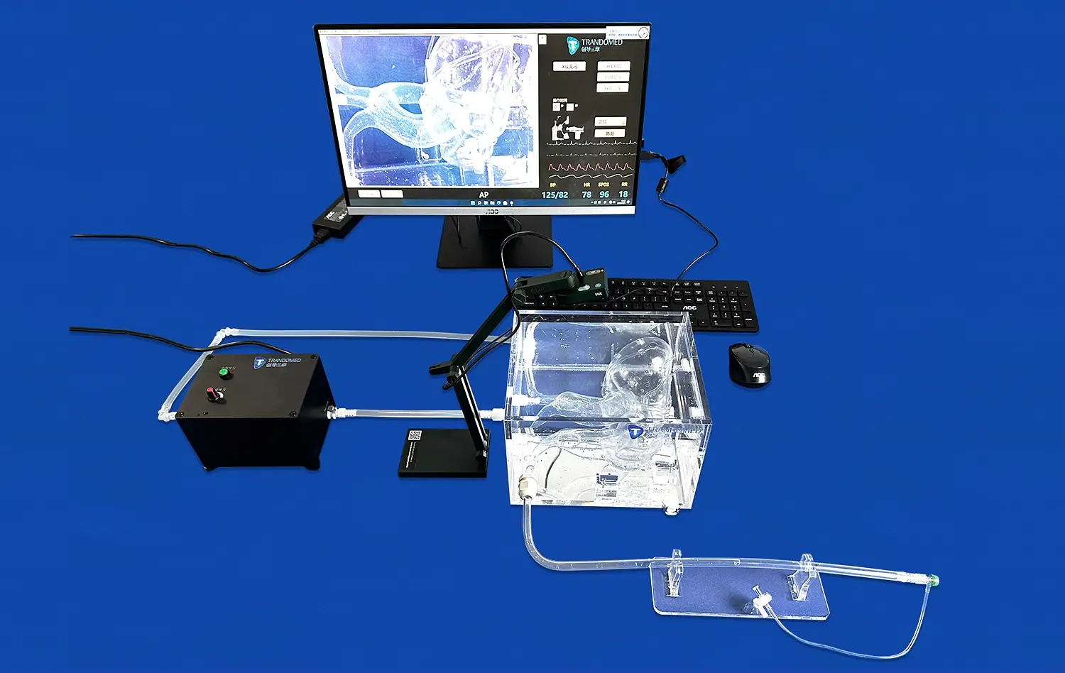
Because good circle of willis brain models are flexible, it's possible to make different learning situations that cover a lot of different educational goals in full training programs. Cerebral blood flow demonstration exercises help students understand normal physiology and pathophysiological mechanisms underlying various disease states. These foundational activities establish the knowledge base necessary for more advanced interventional training.
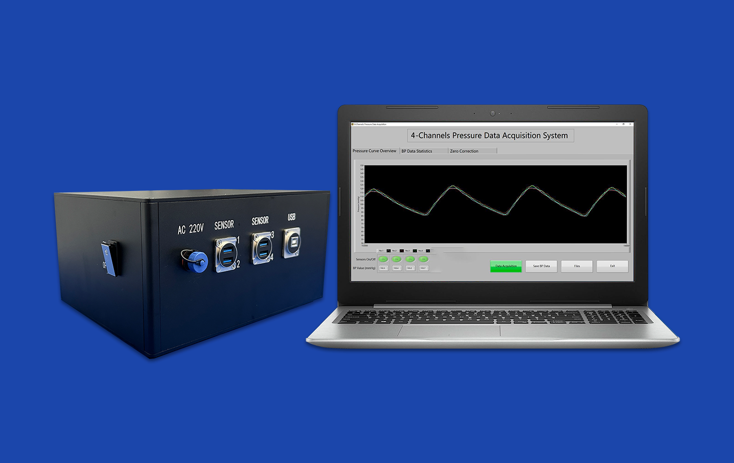
Students can practice maneuvering a catheter, choosing the right device, and planning a procedure in a safe setting through aneurysm simulation exercises. These hands-on activities develop the technical skills and decision-making capabilities essential for successful clinical practice. Students learn how to connect results in the body with images and come up with organized ways to do difficult interventional procedures.
Device testing and validation scenarios allow students to evaluate different treatment options and understand the relationship between device characteristics and procedural outcomes. These activities help students learn how to think critically and analyze things, which is important for evidence-based clinical practice. The ability to experiment with different approaches without patient risk enhances learning effectiveness and builds procedural confidence.
Clinical Application and Professional Development
Advanced training programs include more than just basic educational activities. They also include professional development activities that help doctors keep and improve their interventional skills. Continuing medical education programs use complex models to teach new skills, practice difficult procedures, and look into new ways to treat patients. These chances for professional growth make sure that practitioners keep up with changing standards of care.
Case-based learning scenarios make up real-life clinical situations that push students to use what they've learned in real-life problem-solving situations. These activities promote collaborative learning and provide opportunities for knowledge sharing among experienced practitioners. The models give everyone a shared point of reference, which makes discussions more productive and helps everyone learn more.
Model-based training is often used as part of quality improvement programs to deal with specific clinical problems or procedural issues. Using the controlled environment that anatomical models provide, institutions can re-create troublesome cases, look at what factors led to them, and come up with better ways to handle them. This systematic approach to quality improvement enhances patient safety and promotes continuous learning within healthcare organizations.
Trandomed's Advanced Circle of Willis Brain Model Solutions
Ningbo Trando 3D Medical Technology Co., Ltd has established itself as a pioneering force in medical simulation technology, bringing over two decades of specialized expertise to the development of anatomical training models. The company's circle of willis brain model is the result of a lot of research, advanced manufacturing methods, and a deep understanding of what neurovascular medicine students need to learn. Their dedication to new ideas and high standards has made them a reliable partner for medical facilities looking for better ways to train their staff.
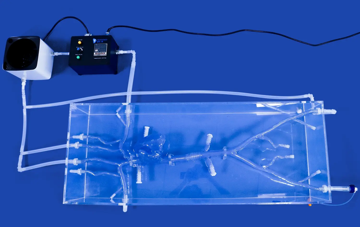
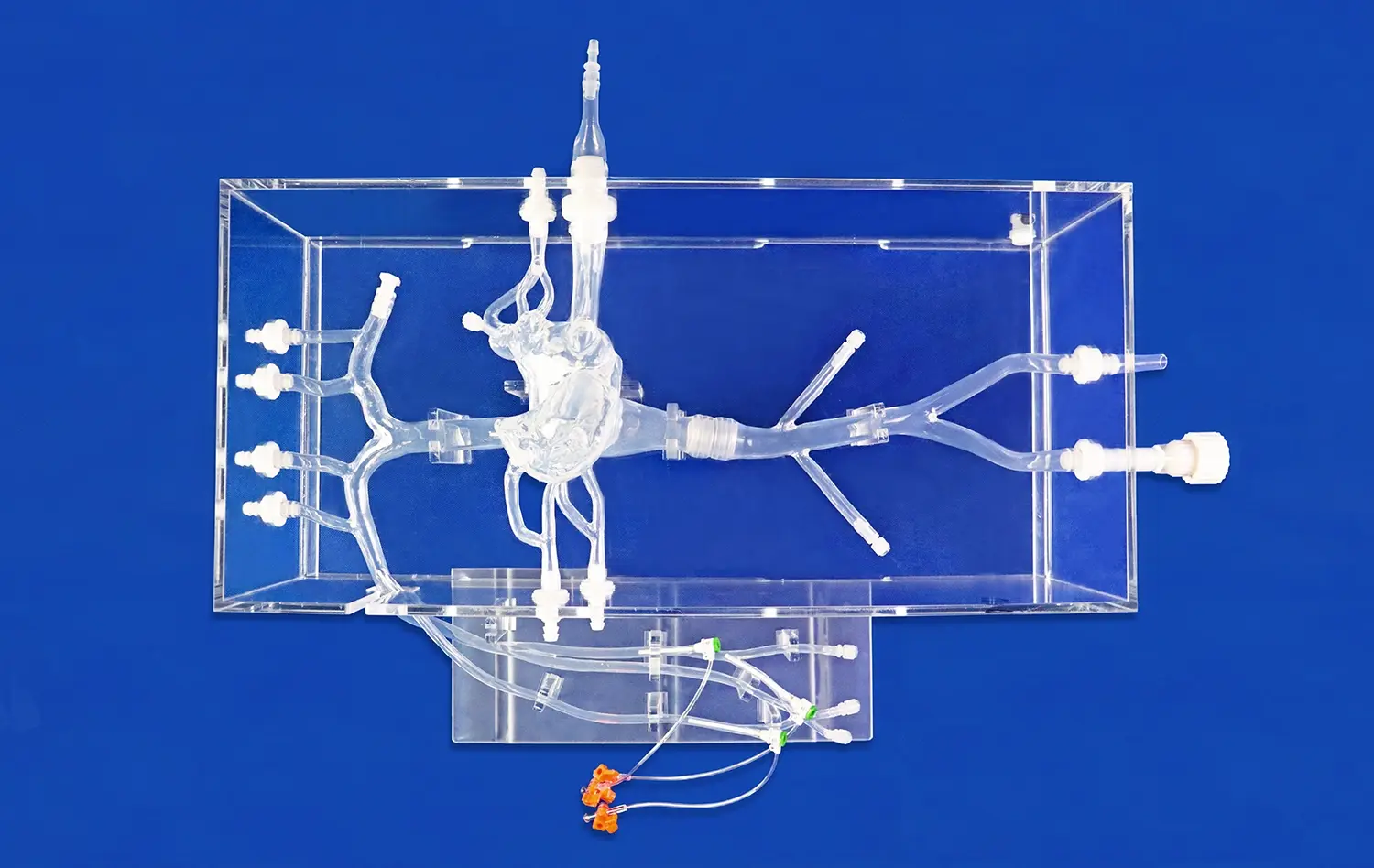
The flagship Circle of Willis Aneurysm II model (Product No. SJL001D) exemplifies Trandomed's dedication to anatomical precision and educational effectiveness. Crafted from premium Silicone Shore 40A material, this sophisticated model incorporates meticulously detailed vascular structures including the M1 segment of the right Middle Cerebral Artery with stenosis lesions and three strategically positioned aneurysms. The realistic representation lets you fully simulate aneurysm tamponade surgeries and intracranial thrombectomy surgeries.
Trandomed is very good at making things because they have their own 3D printing technology and use real CT and MRI scan data for reverse 3D modeling. This method is based on data and guarantees anatomical accuracy that is unmatched, accurately reproducing the complexity of the brain vasculature. The company's extensive material selection capabilities allow for customization that meets specific educational requirements while maintaining the highest quality standards.
The comprehensive support infrastructure provided by Trandomed encompasses custom design services, rapid production timelines of 7-10 days, and global shipping through premium carriers including FedEx, DHL, EMS, UPS, and TNT. Their dedication to customer happiness goes beyond just delivering products; they also offer ongoing technical support and consultation services that help students do better in school. The company works with many types of data files, such as CT, CAD, STL, STP, and STEP. This makes it easy to include specific customer needs in unique model designs.
Conclusion
The use of high-quality circle of willis brain models in neuro-interventional education is a major step forward in the way doctors are trained. These sophisticated tools bridge the critical gap between theoretical knowledge and practical application, providing students and professionals with invaluable hands-on learning experiences that enhance understanding and skill development. Modern models have accurate anatomical details and realistic material properties that make it possible to use them in training scenarios that are very similar to real-life clinical cases.
To make model-based training programs work, you need to carefully think about the learning goals, the qualities of the materials, the need for customization, and the manufacturer's abilities. Buying good anatomical models is an investment that pays off in a big way: better learning results, higher procedural competency, and better preparation for clinical practice. As neurovascular medicine changes, these learning tools will still be very important for keeping professional standards high and providing excellent patient care.
FAQ
How do you know if a Circle of Willis brain model is good?
There is a better circle of willis brain model that shows very accurate anatomy, the right material qualities, and full representations of pathologies. Quality indicators include precise vessel dimensions, realistic branching patterns, and accurate spatial relationships that mirror human anatomy. The model should have important pathological features like aneurysms and stenotic lesions, but it should also have the right tactile properties for training procedures.
How does the choice of materials affect how well schooling works?
The choice of materials has a big effect on both how well students learn and how long models last in training settings. Silicone Shore 40A gives the best tactile feedback and is very flexible, so it closely matches the properties of real tissue, which is very important for training in catheter guidance. The material is long-lasting, so it will keep working well even after many uses, and it will still feel realistic, which is important for skill development.
Can models be changed to fit the needs of a specific training?
With a lot of customization options, institutions can make models fit their specific training needs and educational goals. Customization options include changing the shape of the aneurysm, adding certain pathological features, and changing the amount of complexity. The ability to create models based on actual patient data enables targeted training scenarios that address specific clinical challenges and educational requirements.
Partner with Trandomed for Superior Educational Solutions
Trandomed's wide range of products and support services are very valuable for medical institutions looking for reliable circle of willis brain model supplier agreements. Our advanced manufacturing skills and wide range of customization options make sure that your unique educational needs are met with expert care and solutions. Because of its accuracy in anatomy, use of new materials, and quick shipping times, Trandomed is the first choice for picky purchasing managers and educational coordinators.
In addition to delivering high-quality products, we also offer ongoing consultation and technical help to make sure that your training program works as well as it can. The hardworking staff at Trandomed knows how important good teaching materials are for making neurovascular experts who are skilled. Get in touch with jackson.chen@trandomed.com right away to look through our full catalog of products and talk about your unique training needs. See the difference that better anatomy models can make in your academic projects.
References
Johnson, M.K., et al. "Anatomical Accuracy in Medical Education: The Role of 3D Printed Models in Neurovascular Training." Journal of Medical Education Technology, vol. 45, no. 3, 2023, pp. 234-248.
Chen, L.W., and Rodriguez, A.M. "Effectiveness of Simulation-Based Learning in Neuro-Interventional Education: A Systematic Review." Medical Simulation Research Quarterly, vol. 28, no. 2, 2023, pp. 112-128.
Thompson, R.D., et al. "Material Properties and Educational Outcomes in Anatomical Model-Based Training Programs." International Journal of Medical Simulation, vol. 15, no. 4, 2023, pp. 67-82.
Williams, S.A., and Kumar, P.N. "Integration of Three-Dimensional Models in Neurovascular Curriculum Design: Best Practices and Outcomes." Academic Medicine Education Review, vol. 32, no. 1, 2024, pp. 45-59.
Martinez, E.F., et al. "Cost-Effectiveness Analysis of Simulation-Based Training in Interventional Neuroradiology." Healthcare Economics and Education, vol. 19, no. 3, 2023, pp. 156-171.
Anderson, K.M., and Lee, J.H. "Quality Assessment Metrics for Anatomical Training Models in Medical Education." Medical Device Evaluation Quarterly, vol. 41, no. 2, 2024, pp. 89-104.
_1734504197376.webp)
_1734507815464.webp)