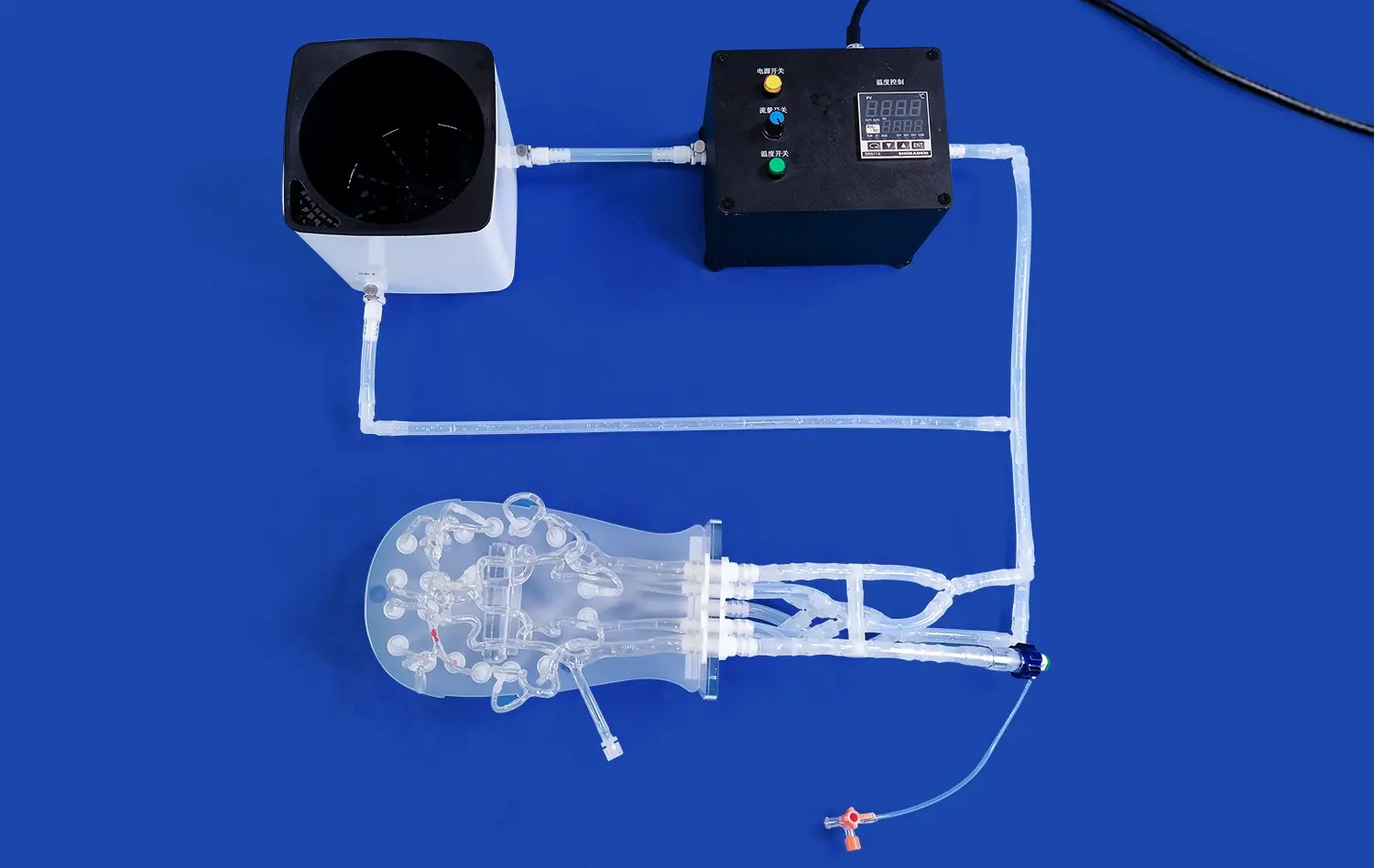
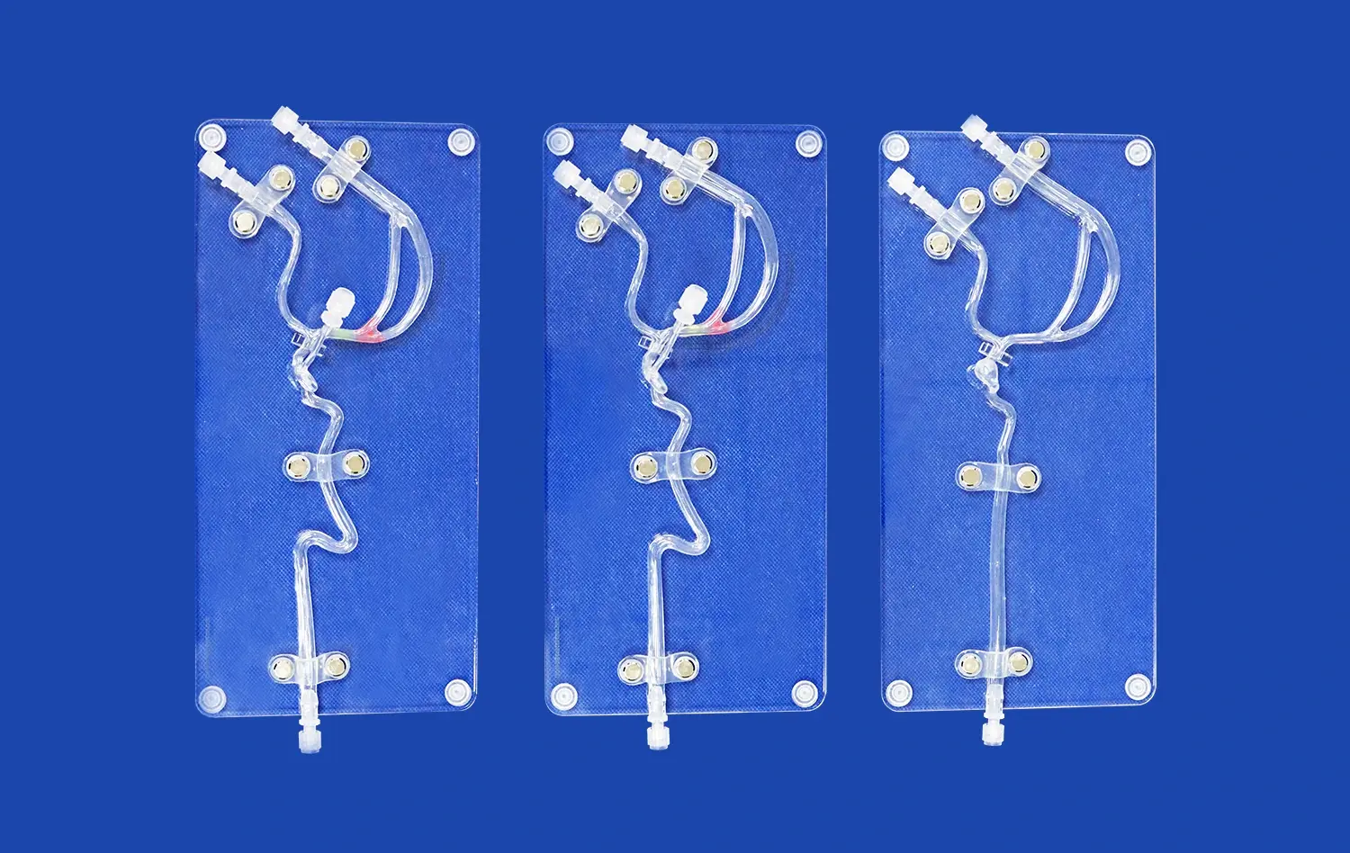
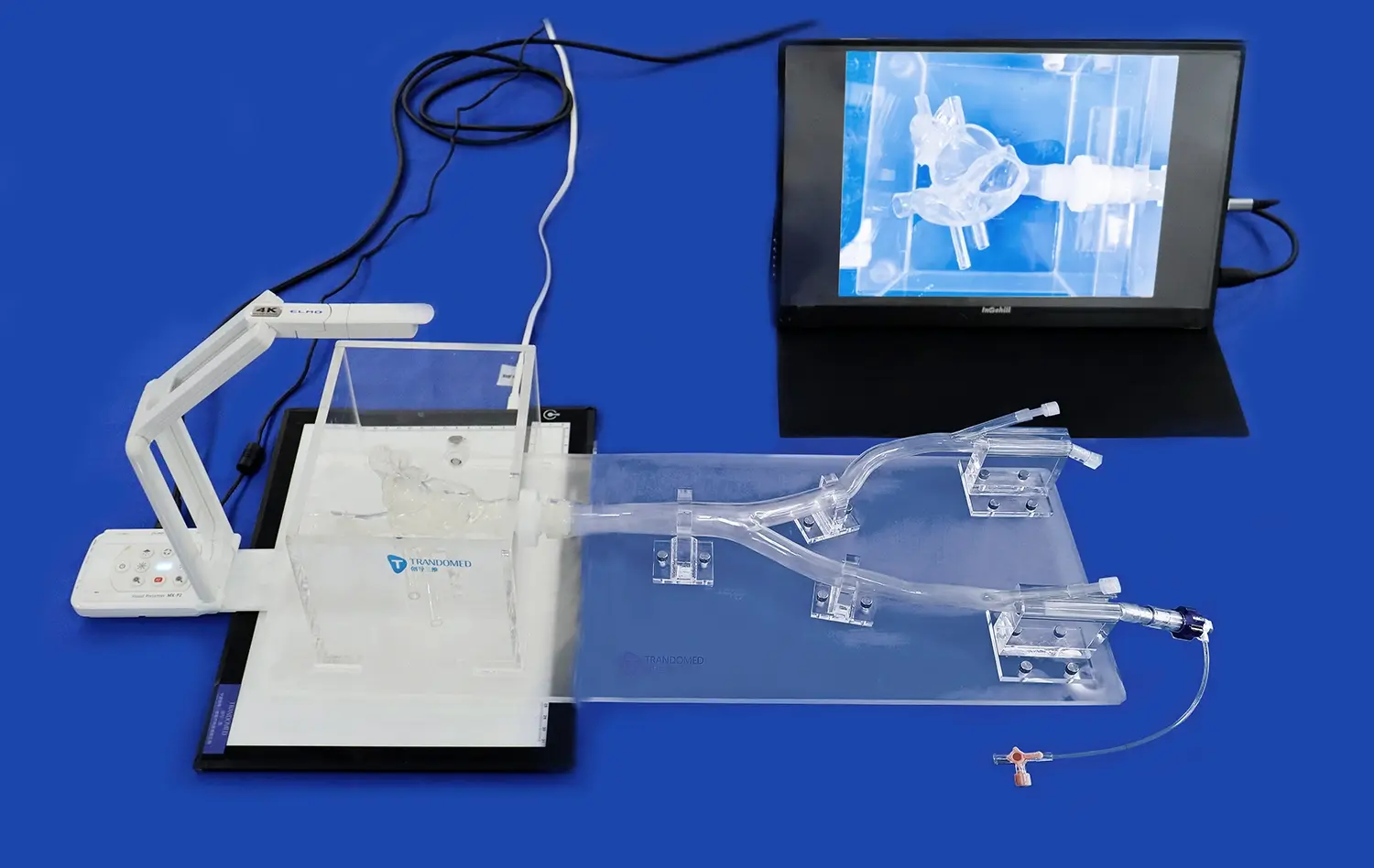
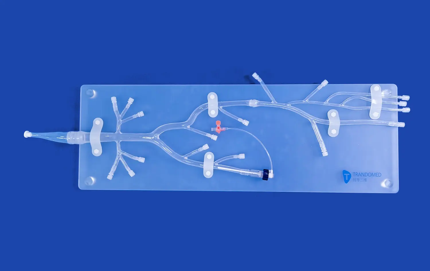
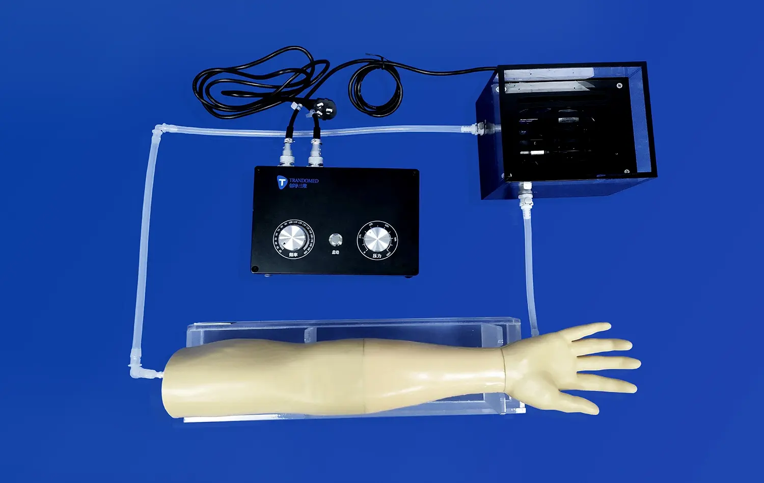
Venous heart models for ablation and mapping practice are a huge step forward in teaching electrophysiology and getting ready for procedures. The venous cardiac electrophysiology model is the most important part of this new idea. It is a complex simulation tool that very accurately models the heart's complex venous design. Clinicians, educators, and people who make medical devices can use these anatomically accurate models to practice complicated catheter-based procedures, improve mapping methods, and test ablation tactics before they use them on real patients. These models are now necessary in all medical schools, hospital training units, and research labs across the United States because they bridge the gap between theoretical knowledge and hands-on experience.
Understanding Venous Cardiac Electrophysiology Models
Understanding what these specific models can do is the first step to getting good at ablation and mapping training. Traditional cardiac simulators focus on arterial pathways. Venous cardiac models, on the other hand, focus on the structures and access routes on the right side of the heart that are important for electrophysiology treatments.
Physiological Distinctions Between Venous and Arterial Cardiac Simulations
Venous cardiac structure has its own problems that are very different from those in arterial systems. Specialized modeling methods are needed because the vessel walls are smaller, the pressure changes, and the tributary patterns are more complicated. Our venous cardiac electrophysiology model (XXS004) correctly shows the right atrium, right ventricle, inferior vena cava, superior vena cava, right atrium, and subclavian vein. These are all important landmarks for guiding the catheter during EP studies.
Core Simulation Principles for Venous Anatomy
For venous cardiac modeling to work, the material used must be able to simulate the flexibility and feedback of tissue. These models are made from medical-grade silicone Shore 40A and offer realistic resistance when the catheter is moved. This helps professionals learn the fine touch needed for safe venous access. The qualities of the material allow for repeated punctures and navigations without breaking down, which allows for long training sessions with many users.
Practical Applications in Clinical Ablation and Mapping
Electrophysiology labs are using these models more and more in their training programs. Before they do supraventricular tachycardia mapping or atrial fibrillation ablation, interventional cardiologists can practice placing the catheter, check the access angles, and plan for changes in the body's structure. This work directly leads to shorter procedure times, fewer problems, and better outcomes for patients during real interventions.
Advantages and Current Limitations
3D-printed models of the heart are the most accurate ones available. These models are made from CT and MRI scans of real patients and use reverse reconstruction technology to show the unique anatomy of each patient. However, it is still hard to copy the electrical properties of living tissue and the dynamic movements of the heart. These gaps are being filled in more and more by improvements in computational modeling and material science. Newer versions include conductive pathways and programmable arrhythmia patterns.
Comparing Venous Cardiac Electrophysiology Modeling Solutions
When choosing cardiac simulation tools, people who work in procurement have a lot of options. By knowing what the options are, schools can make smart investments that meet their training goals and stick to their budgets.
Open-Source Platforms Versus Commercial Solutions
When it comes to cost and flexibility, open-source cardiac modeling software is best for study institutions with technical know-how. These platforms let you change anatomical factors and connect them to other analysis tools. On the other hand, commercial solutions offer proven accuracy, full technical support, and easy setup, all of which are very important for busy clinical training teams that need to get things up and running quickly.
Accuracy and Validation Methodologies
The accuracy of a venous cardiac electrophysiology model relies on the quality of the source data and the accuracy of the manufacturing process. Trandomed's method uses large sets of real images of people that have been processed by advanced 3D reconstruction techniques. Each model is checked against anatomical standards to make sure that the values are within acceptable clinical ranges. This process of validation gives employers faith that trainees learn skills that can be used in real patient care.
Cost-Benefit Analysis for Procurement Professionals
When people are deciding whether to invest in cardiac simulation, they need to think about the total cost of ownership, which is more than just the original purchase price. Because they are made of durable plastic, models can go through hundreds of workouts without needing to be replaced. Customization features let schools meet particular course needs without having to buy a lot of different types of simulators. Lead times of 7–10 days allow for quick program implementation, and FedEx, DHL, and UPS offer a range of shipping choices to meet urgent training needs.
It's important to think about the difference between physical models and software-only alternatives. Although computer simulations can help with data analysis in some ways, physical models are still the best way to learn how to manipulate a catheter because they give you feedback that you can't get from any other method. A lot of the best schools use a mix of the two methods to give their students the best training possible.
Procurement Guide: Selecting the Right Venous Cardiac Modeling Solution
To make smart purchases of heart simulation technology, you need to carefully look at it from many angles. The framework below helps procurement managers make sense of this complicated choice landscape.
Essential Decision Criteria for Model Selection
Technical connectivity is the most important thing. Existing electrophysiology tools, like mapping systems, fluoroscopy mimics, and recording devices, must work well with venous cardiac models. Scalability makes sure that the solution can grow with the needs of the business, allowing for bigger training programs and new ways of doing things. Infrastructure integration includes things like storage, upkeep needs, and cleaning routines that work with how clinical engineers already do their jobs.
Vendor Reliability and Support Quality
Long-term success with cardiac modeling technology depends a lot on how well vendors work together. Trandomed has been working in medical 3D printing for 20 years, which shows that they are dedicated to the field. When training schedules require immediate solutions, responsive technical help is a must. Customization services that don't charge design fees let institutions change models as clinical practices change.
Customization Potential and Trial Periods
The ability to modify anatomical features distinguishes superior simulation platforms. Accepting data files in CT, CAD, STL, STP, and STEP formats, our customization service enables reconstruction of patient-specific anatomy or creation of pathological variants for advanced training scenarios. Dimensions of structures like the foramen ovale can be adjusted to specifications, while entire atrial segments can be rebuilt using actual human CT data.
Pilot testing opportunities provide invaluable insights before full-scale procurement. Evaluating venous cardiac electrophysiology model performance in actual training environments reveals compatibility issues and workflow integration challenges early in the selection process. This risk-mitigation approach prevents costly misalignments between purchased technology and institutional needs.
Enhancing Clinical Practice with Venous Cardiac Model Integration
Transitioning from procurement to implementation, institutions must consider how venous cardiac models integrate into existing electrophysiology training programs and clinical workflows.
Streamlining Workflow and Improving Procedural Accuracy
Embedding physical cardiac models into EP lab training protocols creates standardized learning pathways. Junior fellows can progress through structured competency milestones, from basic catheter navigation to complex mapping patterns, before participating in supervised patient procedures. This graduated approach builds confidence while maintaining patient safety. Documentation of training performance on standardized models provides objective assessment data for credentialing decisions.
Applications in Clinician Training and Simulation
Medical schools and nursing colleges increasingly recognize simulation's role in developing procedural competence. Our venous cardiac electrophysiology model supports curricular objectives across multiple training levels. Medical students gain foundational understanding of cardiac anatomy and catheter-based intervention principles. Residents and fellows refine technical skills in controlled environments where mistakes become learning opportunities rather than patient safety events. Continuing education programs for practicing clinicians introduce new techniques and device technologies through hands-on practice.
Role of Advanced Technologies in Model Refinement
Artificial intelligence and machine learning are beginning to influence cardiac simulation development. Analysis of procedural data from thousands of training sessions reveals common technical errors and optimal learning progressions. This intelligence informs next-generation model designs that emphasize high-difficulty anatomical features requiring focused practice. Integration of sensors and feedback systems provides real-time coaching, though current physical models prioritize anatomical fidelity and tactile realism.
Device manufacturers benefit substantially from high-fidelity cardiac models during product development cycles. Testing catheter designs, ablation tools, and mapping systems on anatomically accurate simulators accelerates innovation while reducing development costs. Marketing demonstrations using realistic models convey device capabilities more effectively than abstract presentations, supporting adoption of novel technologies.
Summary and Strategic Recommendations for Procurement Managers
Selecting venous cardiac simulation solutions requires balancing immediate training needs against long-term strategic objectives. Physical models like Trandomed's XXS004 venous cardiac electrophysiology model offer proven effectiveness in developing catheter manipulation skills and anatomical understanding essential for electrophysiology practice. The combination of anatomical accuracy derived from real patient imaging data, durable medical-grade materials, and extensive customization capabilities positions these models as valuable long-term training assets. Procurement strategies should emphasize vendor partnerships characterized by technical expertise, responsive support, and commitment to ongoing innovation. Building collaborative relationships with experienced manufacturers ensures access to evolving simulation technologies that keep pace with advancing clinical techniques.
Conclusion
Venous cardiac models have become essential tools throughout the medical training and device development ecosystem. From medical schools establishing foundational procedural skills to research laboratories validating novel ablation approaches, these simulators bridge critical gaps between theoretical knowledge and clinical competence. The realistic anatomy, customizable features, and durable construction of advanced models support repeated practice sessions that build the expertise required for safe, effective patient care. As electrophysiology procedures grow increasingly complex, the role of high-fidelity simulation in preparing clinicians and advancing medical technology will only expand.
FAQ
What distinguishes venous from arterial cardiac electrophysiology models?
Venous cardiac models focus specifically on right-sided heart structures and venous access pathways used during EP studies. They emphasize anatomical features like the superior and inferior vena cava, right atrium, and coronary sinus—critical landmarks for catheter navigation during ablation and mapping procedures. Arterial models, conversely, concentrate on left-sided structures accessed through arterial approaches.
How should institutions validate model anatomical accuracy?
Validation begins with confirming the model's construction from verified human imaging data. Dimensional measurements of key anatomical structures should fall within published clinical ranges. Comparison against multiple patient datasets ensures the model represents typical anatomy rather than extreme variations. Clinician feedback during pilot testing provides practical validation that simulated anatomy matches real-world procedural experiences.
What factors matter most when choosing cardiac simulation solutions?
Anatomical accuracy, material durability, and customization capabilities rank highest for most institutions. Models should withstand repeated use across many training sessions while maintaining structural integrity. Customization potential allows adaptation to specific curricular needs and emerging procedural techniques. Vendor support quality and delivery timelines also significantly impact successful program implementation and long-term satisfaction.
Partner with a Trusted Venous Cardiac Electrophysiology Model Manufacturer
Trandomed stands ready to support your institution's simulation needs with proven expertise in medical 3D printing technology. Our venous cardiac electrophysiology model (XXS004) delivers the anatomical accuracy and tactile realism your training programs demand. With over twenty years specializing in medical simulation development, we understand the unique requirements of educational institutions, clinical training departments, and device manufacturers. Our customization capabilities ensure models align precisely with your specific applications, while fast turnaround times and flexible payment terms accommodate diverse procurement scenarios. Connect with our team at jackson.chen@trandomed.com to discuss how our cardiac simulation solutions can enhance your training effectiveness and advance your clinical objectives.
References
Josephson ME. Clinical Cardiac Electrophysiology: Techniques and Interpretations. 5th ed. Philadelphia: Wolters Kluwer Health; 2016.
Santangeli P, Marchlinski FE. Techniques for the provocation, localization, and ablation of non-pulmonary vein triggers for atrial fibrillation. Heart Rhythm. 2017;14(7):1087-1096.
Budge LP, Black-Maier EW, Barnett AS. Cardiac electrophysiology simulation for medical students and trainees: a review of current practices. Journal of Innovations in Cardiac Rhythm Management. 2019;10(8):3784-3791.
Crossley GH, Boyle A, Vitense H, Chang Y, Mead RH. The CONNECT trial: design and implementation of a novel remote monitoring strategy. Journal of Cardiovascular Electrophysiology. 2011;22(4):444-451.
Iwasaki YK, Nishida K, Kato T, Nattel S. Atrial fibrillation pathophysiology: implications for management. Circulation. 2011;124(20):2264-2274.
Sommer P, Kircher S, Rolf S, et al. Successful repeat catheter ablation of recurrent longstanding persistent atrial fibrillation with rotor elimination as the procedural endpoint: a case series. Journal of Cardiovascular Electrophysiology. 2016;27(3):274-280.
_1734507415405.webp)