Demonstrating Detailed Anatomy of Peripheral Abdominal Arteries
Precise Replication of Vascular Structures
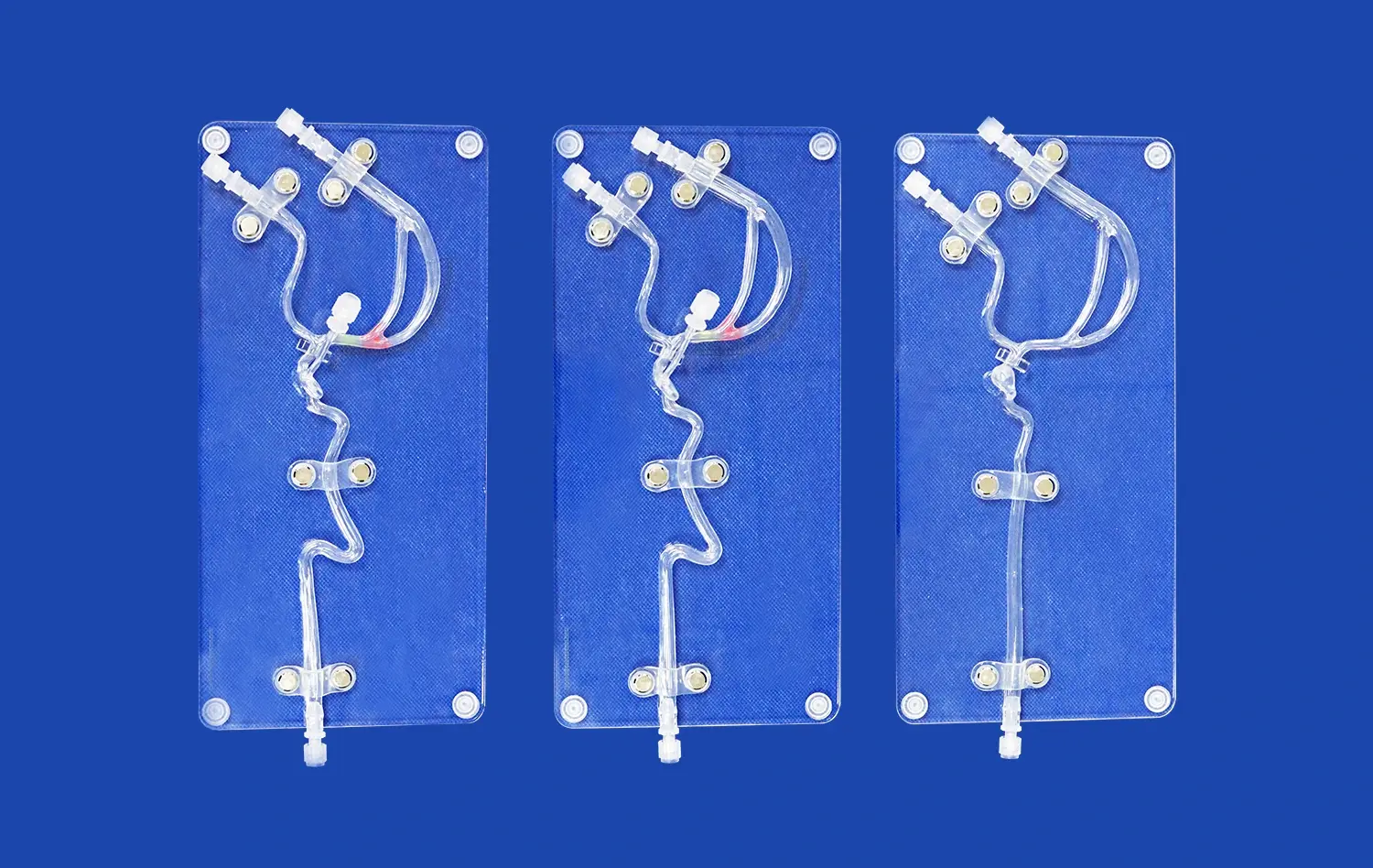
The abdominal vascular model excels in its ability to precisely replicate the intricate network of peripheral abdominal arteries. Using advanced 3D printing technology, these models capture even the finest details of vascular anatomy, including vessel diameter, branching patterns, and spatial relationships. This level of precision is crucial for medical professionals and students to gain a comprehensive understanding of the abdominal vascular system.
The model typically includes accurate representations of key vessels such as the abdominal aorta, celiac trunk, hepatic artery, splenic artery, renal arteries, and iliac arteries. By visualizing these structures in a three-dimensional format, learners can develop a more intuitive grasp of vascular anatomy compared to traditional two-dimensional images or textbook illustrations.
Visualization of Anatomical Variations
One of the significant advantages of using an abdominal vascular model is its ability to demonstrate anatomical variations. In real-life clinical scenarios, physicians often encounter patients with unique vascular anatomies that deviate from the textbook norm. The model can be customized to showcase these variations, preparing medical professionals for the diverse range of anatomical structures they may encounter in practice.
For instance, the model can depict variations in the origin and branching patterns of the celiac trunk, superior mesenteric artery, or renal arteries. This feature is particularly valuable for interventional radiologists and vascular surgeons who need to navigate complex anatomies during procedures.
Enhanced Spatial Understanding
The three-dimensional nature of the abdominal vascular model significantly enhances spatial understanding of peripheral arteries. Unlike flat images or diagrams, the model allows learners to observe the vascular structures from multiple angles and perspectives. This multidimensional view is crucial for grasping the spatial relationships between different arteries and surrounding organs.
For example, the model can clearly demonstrate how the renal arteries relate to the kidneys, or how the hepatic artery interacts with the liver and gallbladder. This spatial comprehension is invaluable for planning surgical approaches, understanding potential complications, and visualizing the path of interventional devices through the vascular system.
Teaching Complex Vascular Pathways with High Fidelity
Realistic Simulation of Blood Flow
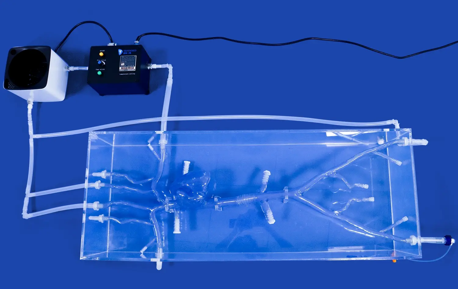
Advanced abdominal vascular models often incorporate features that simulate blood flow, adding another layer of realism to the learning experience. Some models are designed with hollow vessels that can be filled with colored fluids to mimic blood flow. This dynamic aspect allows learners to observe how blood moves through the peripheral arteries, enhancing their understanding of hemodynamics in the abdominal region.
The ability to simulate blood flow is particularly beneficial for demonstrating conditions such as stenosis or aneurysms. By adjusting the flow rate or introducing obstructions, instructors can illustrate how these pathologies affect blood circulation in peripheral arteries, providing valuable insights into disease progression and potential complications.
Integration of Pathological Conditions
High-fidelity abdominal vascular models can be designed to incorporate various pathological conditions, offering a unique opportunity to study abnormalities in a controlled environment. These models can showcase common vascular diseases such as atherosclerosis, arterial dissection, or abdominal aortic aneurysms.
By integrating these pathologies into the model, medical educators can provide hands-on demonstrations of how diseases affect the structure and function of peripheral arteries. This approach allows learners to visualize the progression of vascular conditions and understand their impact on surrounding tissues, fostering a more comprehensive understanding of abdominal vascular pathology.
Multimodal Learning Opportunities
The versatility of the abdominal vascular model enables multimodal learning experiences, catering to diverse learning styles and educational objectives. These models can be used in conjunction with other teaching methods to create a comprehensive learning environment.
For instance, the model can be paired with imaging studies such as CT angiograms or MRI scans, allowing learners to correlate the three-dimensional structure with two-dimensional imaging findings. This integration of multiple learning modalities enhances the overall educational experience and helps bridge the gap between theoretical knowledge and practical application in clinical settings.
Improving Understanding of Peripheral Artery Interventions
Hands-on Practice for Interventional Procedures
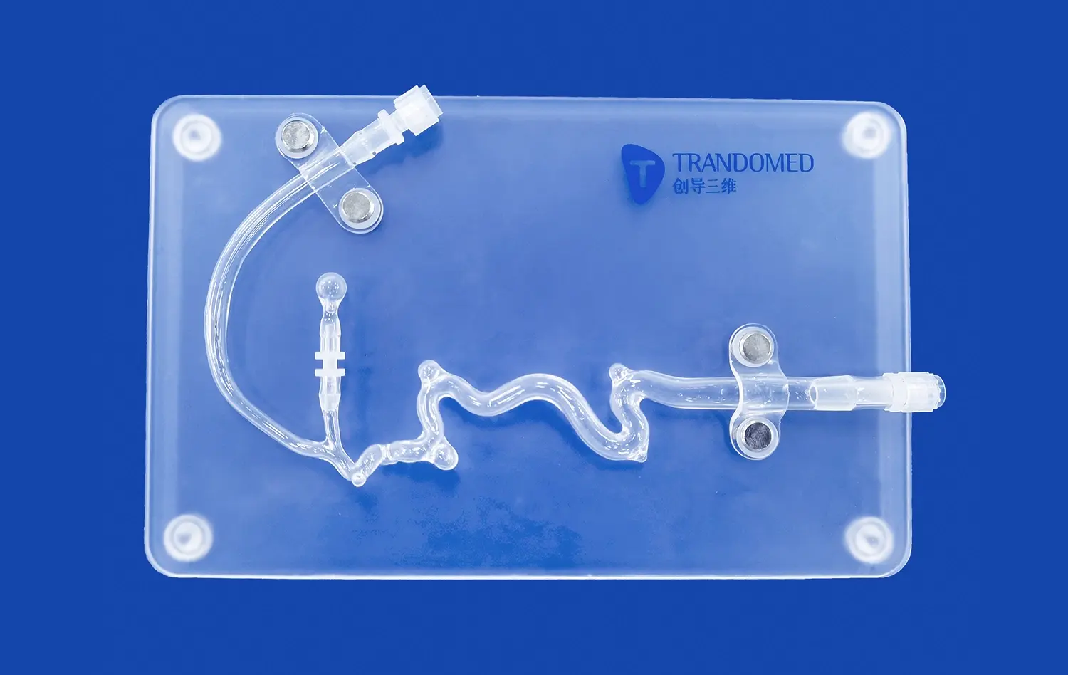
One of the most significant advantages of the abdominal vascular model is its ability to provide a platform for hands-on practice of interventional procedures. These models are often designed with materials that simulate the properties of human tissue, allowing learners to practice techniques such as catheter insertion, guidewire navigation, and stent deployment in a risk-free environment.
This practical experience is invaluable for developing the fine motor skills and spatial awareness required for successful peripheral artery interventions. Learners can repeatedly practice complex procedures, refining their techniques without the pressure of working on actual patients. This approach not only improves procedural competence but also enhances confidence and reduces the learning curve associated with interventional techniques.
Demonstration of Interventional Approaches
The abdominal vascular model serves as an excellent tool for demonstrating various interventional approaches used in peripheral artery procedures. Instructors can use the model to illustrate different access points, such as femoral or radial approaches, and demonstrate how to navigate through the vascular system to reach specific target areas.
Moreover, the model can be used to showcase different interventional techniques, such as balloon angioplasty, stenting, or atherectomy. By visualizing these procedures on the model, learners can better understand the principles behind each technique, the challenges associated with different anatomical locations, and the potential risks and complications that may arise during interventions.
Assessment and Skill Evaluation
The abdominal vascular model provides an objective means of assessing and evaluating interventional skills. Educators can use the model to create standardized scenarios for testing learners' proficiency in performing peripheral artery interventions. This assessment can include factors such as procedural time, accuracy of device placement, and ability to navigate complex vascular pathways.
Furthermore, the model allows for the implementation of performance metrics and feedback systems. For instance, some advanced models may incorporate sensors or imaging capabilities that can track the movement of interventional devices within the vascular system. This data can be used to provide detailed feedback on technique, helping learners identify areas for improvement and track their progress over time.
Conclusion
The abdominal vascular model has proven to be an invaluable asset in peripheral artery demonstrations, significantly enhancing medical education and training. Its ability to provide detailed anatomical representations, simulate complex vascular pathways, and facilitate hands-on practice of interventional procedures makes it an essential tool for healthcare professionals. By bridging the gap between theoretical knowledge and practical application, these models contribute to improved patient outcomes and safer medical practices. As technology continues to advance, we can expect even more sophisticated and realistic abdominal vascular models to further revolutionize the field of vascular medicine and interventional radiology.
Contact Us
Experience the cutting-edge technology and unparalleled realism of Trandomed's abdominal vascular models. As a leading manufacturer and supplier of 3D printed medical simulators, we offer customizable solutions to meet your specific training and demonstration needs. Elevate your medical education and interventional skills with our high-fidelity models. Contact us today at jackson.chen@trandomed.com to learn more about our products and how we can support your educational or clinical objectives.
_1736216292718.webp)
_1734507205192.webp)
_1734507815464.webp)