Compatibility with Advanced Imaging Modalities
Optimized Material Properties for Imaging
The pulmonary vein model is crafted from specially formulated silicone with a Shore 40A hardness, carefully selected to mimic the radiodensity and magnetic properties of human tissue. This material composition ensures optimal visibility and contrast during CTA, DSA, and MRA procedures, allowing for clear differentiation between vascular structures and surrounding tissues. The model's ability to replicate tissue-like properties under various imaging conditions contributes to more accurate and reliable study results.
Enhanced Contrast Agent Dynamics
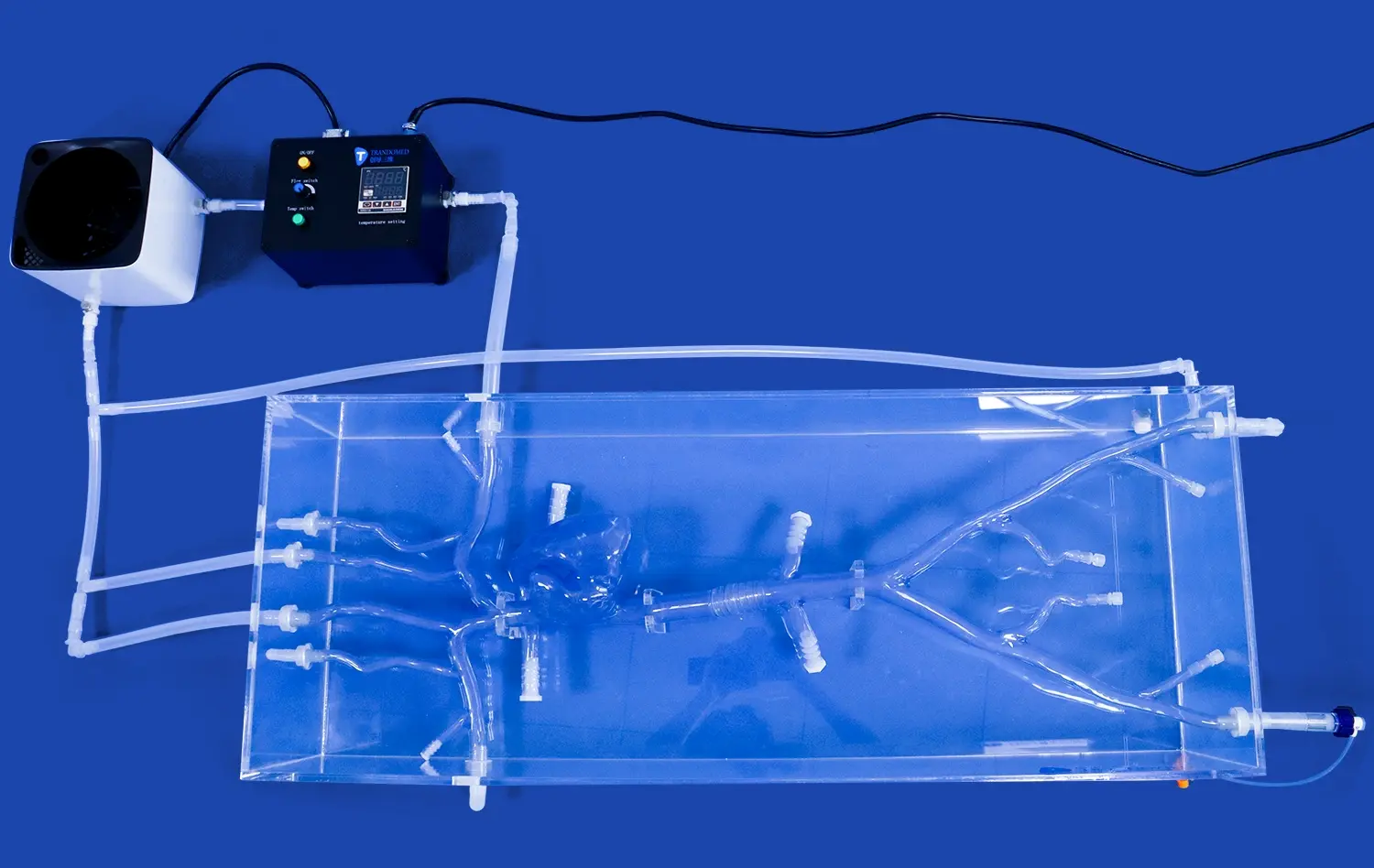
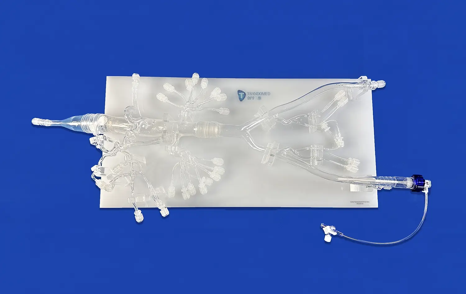
Designed with precision, the pulmonary vein model features strategically placed holes in the left and right atrium, facilitating the introduction of contrast agents. This design element allows for realistic simulation of contrast dynamics within the pulmonary venous system. Researchers and clinicians can observe the flow and distribution of contrast media, mimicking actual patient scenarios. This capability is particularly valuable for assessing the effectiveness of new contrast agents or optimizing imaging protocols for pulmonary vein visualization.
Multi-modality Imaging Support
The versatility of the pulmonary vein model extends beyond CTA, DSA, and MRA, supporting additional imaging techniques such as Optical Coherence Tomography (OCT) and Particle Image Velocimetry (PIV). This multi-modality compatibility enables comprehensive vascular assessments, combining the strengths of different imaging technologies to provide a more complete picture of pulmonary vein anatomy and function. Researchers can leverage this feature to conduct comparative studies across imaging modalities, enhancing diagnostic accuracy and treatment planning.
Detailed Visualization of Blood Flow Patterns
Realistic Vascular Geometry
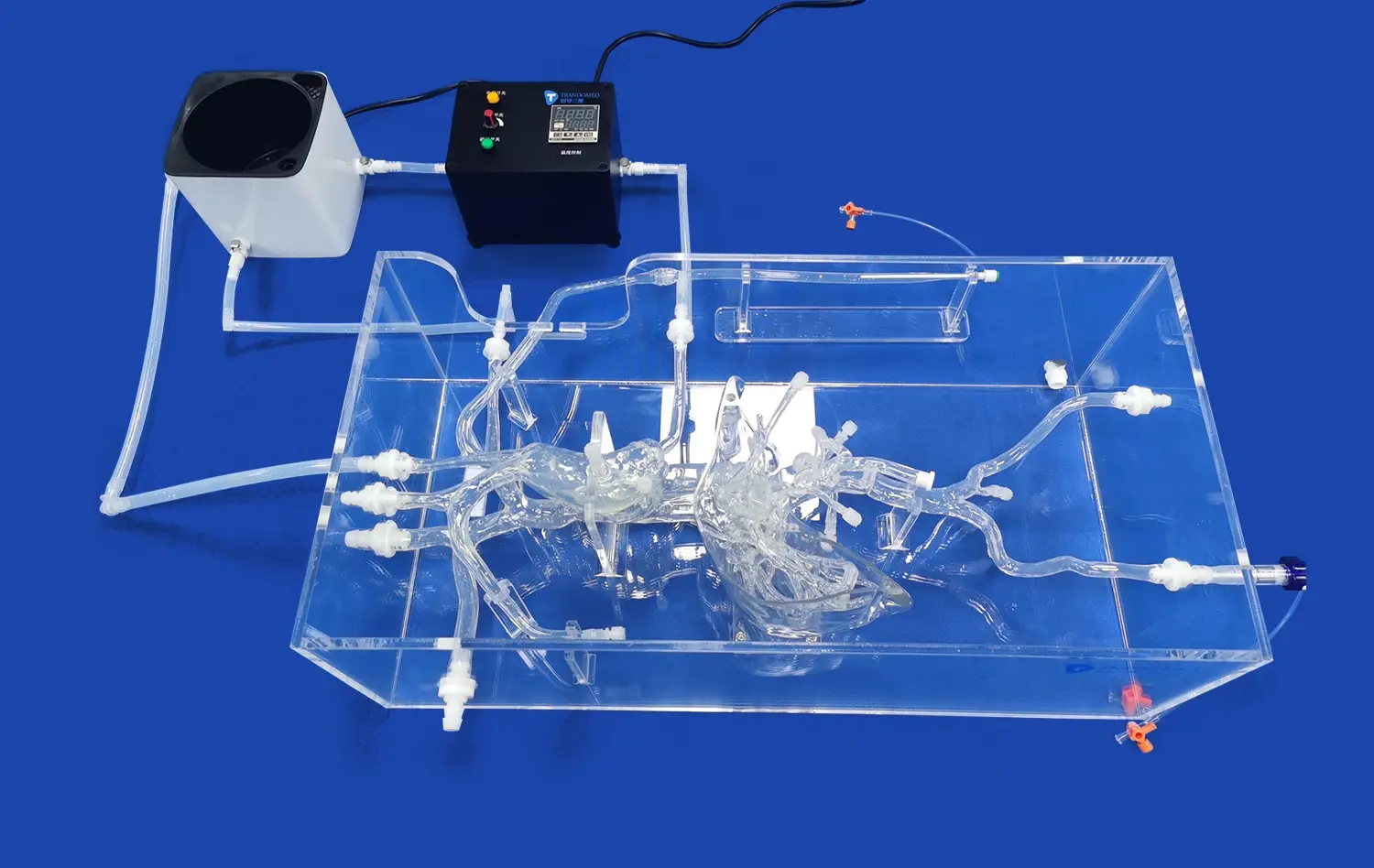
The pulmonary vein model's design is based on real human CT and MRI data, refined through advanced reverse 3D reconstruction technology. This approach ensures that the model accurately represents the complex geometry of the pulmonary venous system, including the four main branches of the pulmonary veins. The precise replication of vascular curvatures, bifurcations, and diameter variations allows for the study of intricate blood flow patterns that closely mimic in vivo conditions.
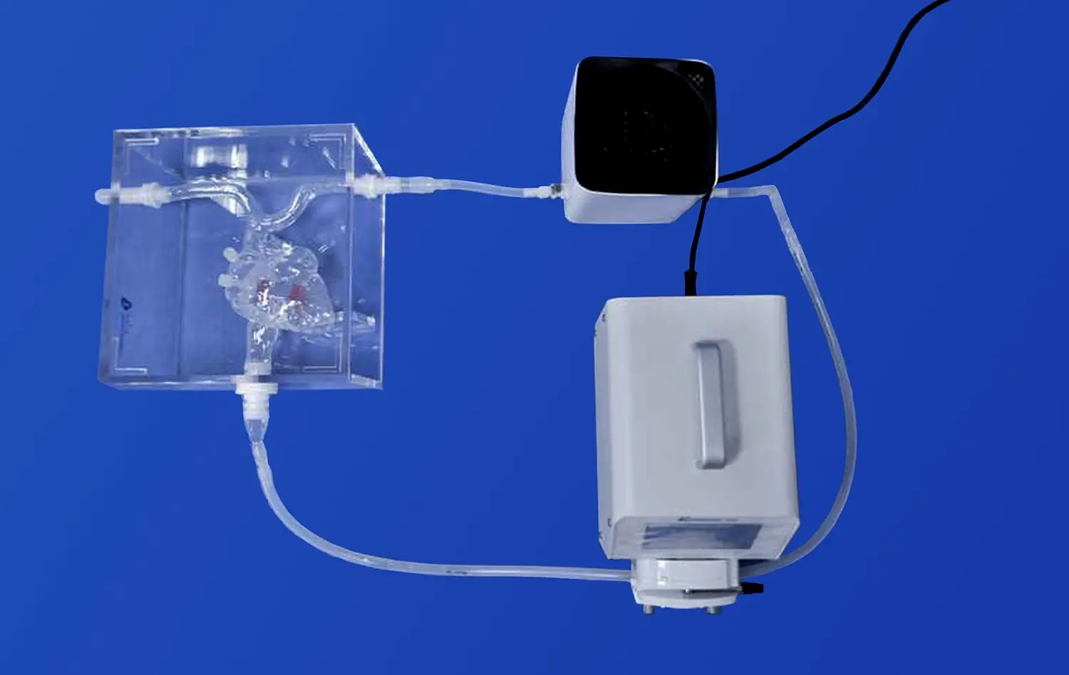
Flow Dynamics Simulation
Equipped with custom transparent pagoda connectors, the pulmonary vein model facilitates the controlled introduction of fluid to simulate blood flow. This feature, combined with the model's anatomically correct structure, enables researchers to observe and analyze complex flow dynamics within the pulmonary veins. Studies can focus on areas of interest such as flow turbulence, stagnation points, and velocity profiles, providing valuable insights into potential thrombosis risk factors or the hemodynamic effects of various pathological conditions.
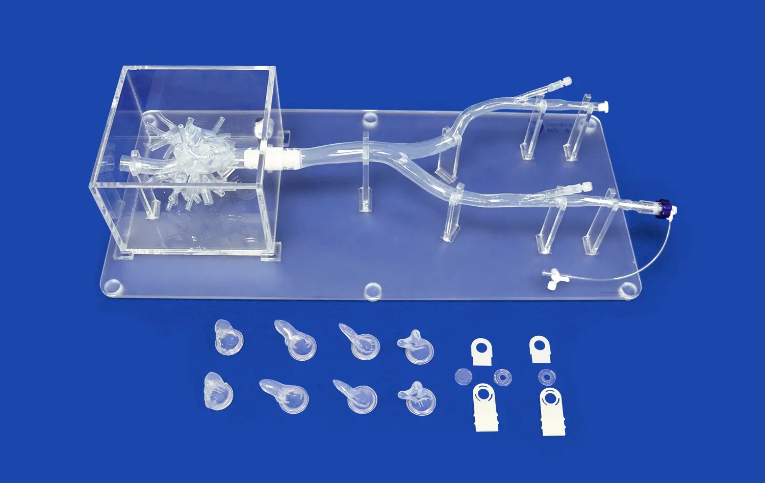
Pathological Variant Integration
The modular design of the pulmonary vein model allows for the integration of various pathological variants. Researchers can incorporate features such as stenosis, aneurysms, or atrial septal defects to study their effects on blood flow patterns. This capability is particularly valuable for understanding how structural abnormalities influence hemodynamics and for developing targeted treatment strategies. The ability to visualize these flow alterations through advanced imaging techniques provides a powerful tool for both educational and research purposes.
Supporting Accurate Device Performance Assessment
Realistic Device-Tissue Interaction
The silicone composition of the pulmonary vein model, with its carefully calibrated Shore 40A hardness, closely mimics the mechanical properties of human vascular tissue. This similarity allows for authentic simulation of device-tissue interactions during interventional procedures. Researchers and device manufacturers can assess the performance of guidewires, catheters, balloons, and stents under conditions that closely resemble clinical scenarios. The model's ability to replicate tissue response to device manipulation provides valuable insights into factors such as device trackability, pushability, and overall maneuverability.
Customizable Pathological Features
Trandomed's pulmonary vein model offers extensive customization options, allowing researchers to incorporate specific pathological features relevant to their studies. Whether it's the addition of pulmonary vein stenosis, left atrial appendage anomalies, or variations in vascular tortuosity, the model can be tailored to meet unique research requirements. This customization capability enables targeted evaluation of device performance in challenging anatomical scenarios, supporting the development and refinement of specialized interventional tools and techniques.
Quantitative Performance Metrics
The integration of advanced imaging capabilities with the pulmonary vein model facilitates the collection of quantitative performance metrics for medical devices. Researchers can utilize techniques such as CTA, DSA, and MRA to assess device deployment accuracy, expansion characteristics, and positional stability under simulated physiological conditions. The model's transparent design elements further enhance visibility, allowing for direct observation and measurement of device behavior. This comprehensive approach to performance assessment contributes to the development of safer, more effective interventional devices for pulmonary vein-related procedures.
Conclusion
The pulmonary vein model emerges as an indispensable tool for CTA, DSA, and MRA studies, offering unparalleled advantages in anatomical accuracy, imaging compatibility, and versatility. Its ability to provide detailed visualization of blood flow patterns and support precise device performance assessments makes it an invaluable asset in cardiovascular research and medical education. By bridging the gap between clinical practice and experimental research, this advanced simulation tool continues to drive innovation in pulmonary vein interventions and imaging techniques, ultimately contributing to improved patient outcomes in cardiovascular care.
Contact US
Elevate your cardiovascular research and training capabilities with Trandomed's state-of-the-art pulmonary vein models. As a leading 3D printed medical simulators manufacturer and supplier, we offer customizable solutions to meet your specific needs. Experience the benefits of our advanced simulation technology, backed by over 20 years of expertise in medical model design and production. Contact us today at jackson.chen@trandomed.com to explore how our pulmonary vein models can enhance your institution's research and educational programs.
_1734504197376.webp)