How PIV Techniques Benefit from Controlled Vascular Models?
Enhanced Flow Visualization
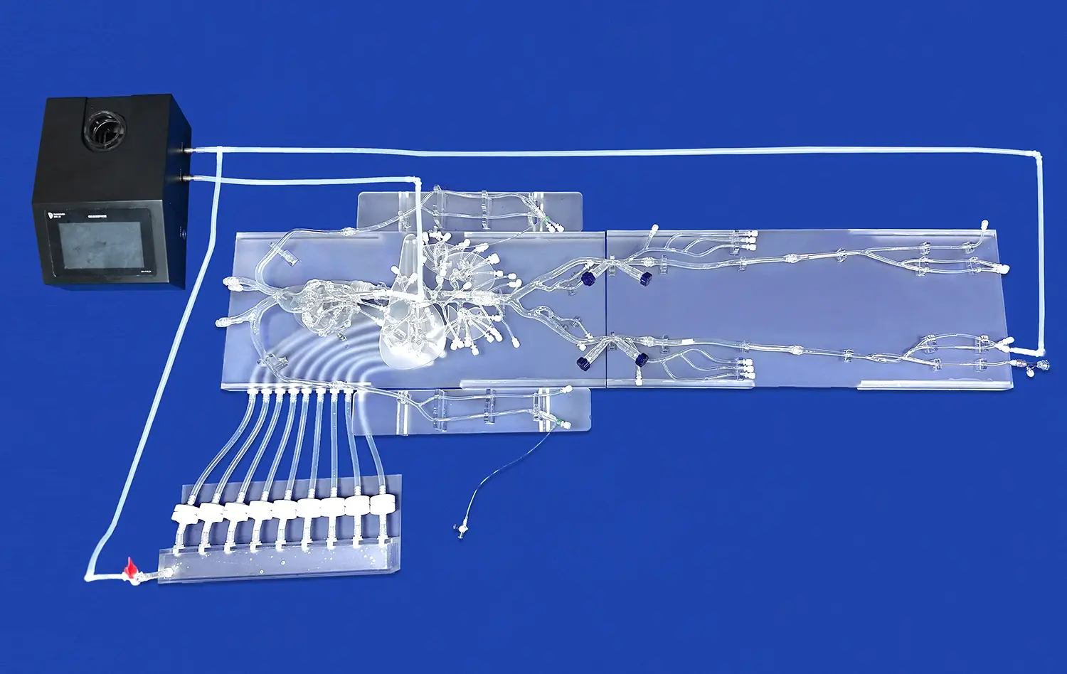
Abdominal vascular models provide a clear advantage in PIV studies by offering superior flow visualization capabilities. The transparent nature of these silicone replicas allows researchers to observe and capture intricate flow patterns that would otherwise be challenging to discern in vivo. This enhanced visibility enables the identification of subtle flow phenomena, such as recirculation zones, vortex formation, and boundary layer separation, which are critical in understanding the progression of vascular diseases.
Precise Geometry Control
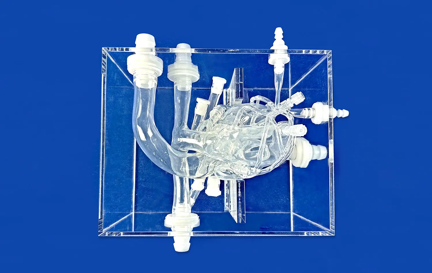
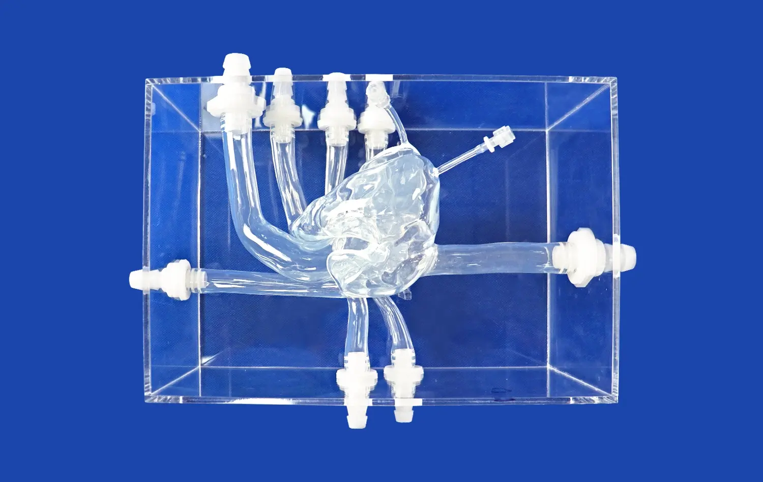
One of the key benefits of using abdominal vascular models in PIV studies is the ability to control and manipulate the vessel geometry with high precision. Researchers can create models that accurately represent specific patient anatomies or idealized vessel configurations, allowing for systematic investigations of how geometric variations influence flow dynamics. This level of control is particularly valuable when studying the effects of arterial stenoses, aneurysms, or complex bifurcations on blood flow patterns.
Reproducibility and Standardization
Controlled vascular models significantly enhance the reproducibility of PIV experiments. By using standardized model geometries and flow conditions, researchers can conduct multiple trials with consistent results, facilitating comparative studies and meta-analyses. This reproducibility is essential for validating computational fluid dynamics (CFD) simulations and developing robust protocols for clinical applications of PIV techniques in vascular diagnostics.
Capturing Detailed Flow Patterns in Complex Arterial Networks
High-Resolution Velocity Mapping
Abdominal vascular models enable high-resolution velocity mapping of blood flow in complex arterial networks. By employing advanced PIV techniques, researchers can obtain detailed spatiotemporal information on flow velocities throughout the model. This high-fidelity data is crucial for understanding the distribution of wall shear stress, which plays a significant role in the development of atherosclerosis and other vascular pathologies. The ability to capture these intricate flow patterns with precision allows for more accurate assessment of potential risk factors and intervention strategies.
Investigating Flow Separation and Recirculation
One of the most valuable aspects of using abdominal vascular models in PIV studies is the ability to investigate flow separation and recirculation phenomena. These flow characteristics are often associated with the development of atherosclerotic plaques and aneurysms. By creating models that replicate specific arterial geometries, such as the abdominal aorta with its branching vessels, researchers can visualize and quantify regions of disturbed flow. This information is invaluable for understanding the biomechanical factors contributing to vascular disease progression and for developing targeted therapeutic approaches.
Analyzing Pulsatile Flow Dynamics
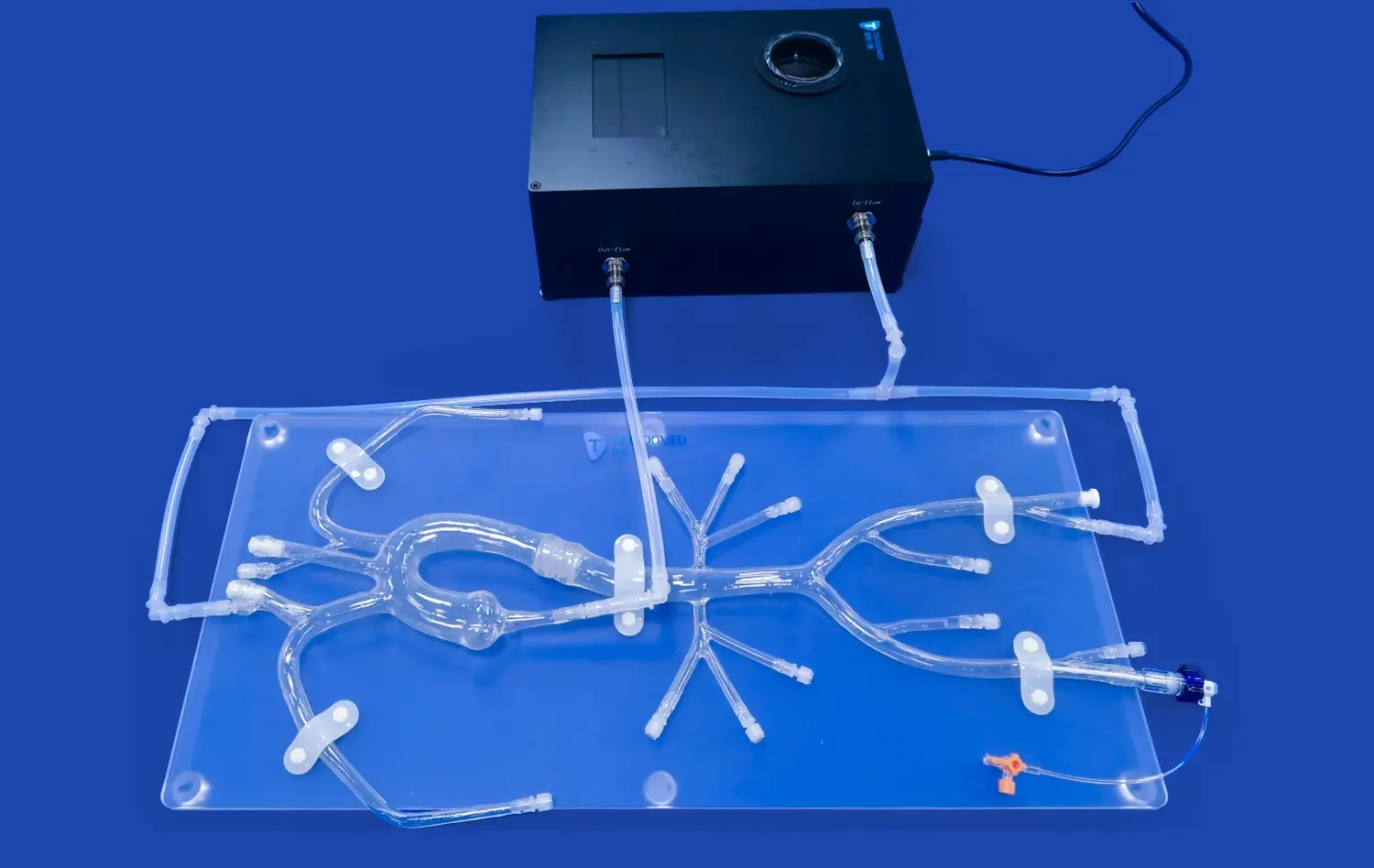
Abdominal vascular models allow for the analysis of pulsatile flow dynamics, which is essential for accurately representing physiological blood flow conditions. By incorporating pulsatile flow generators, researchers can simulate the cyclic nature of arterial blood flow and study its effects on flow patterns and wall shear stress distribution. This capability is particularly important when investigating the hemodynamics of conditions such as abdominal aortic aneurysms, where the pulsatile nature of blood flow plays a crucial role in disease progression and potential rupture risk.
Advancing Research Accuracy with Reproducible Vascular Simulations
Validation of Computational Models
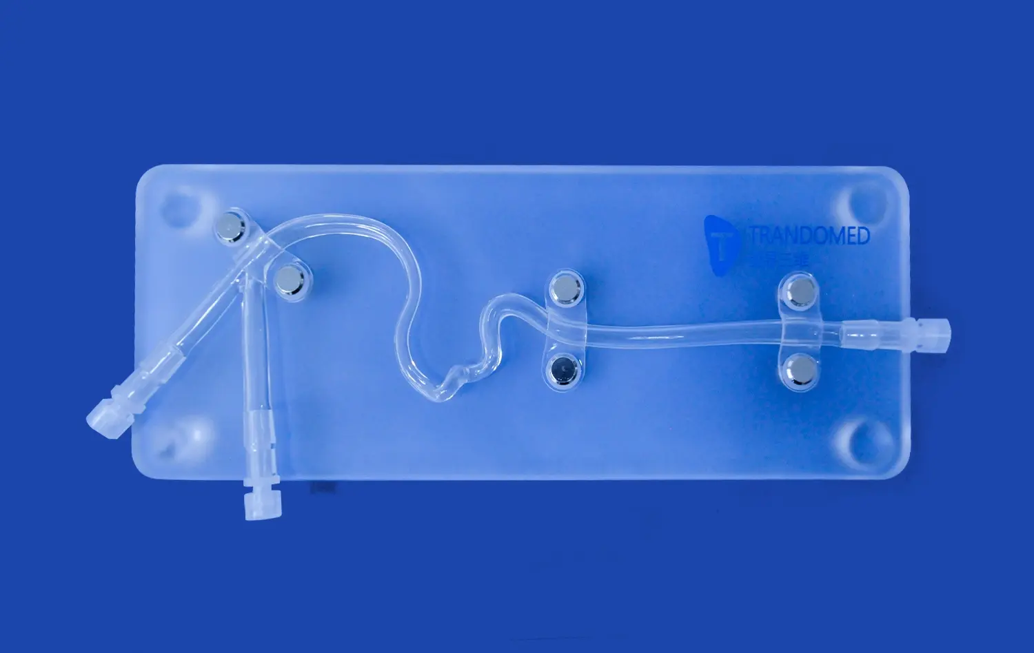
Abdominal vascular models play a vital role in validating computational fluid dynamics (CFD) simulations. By comparing PIV measurements obtained from physical models with CFD predictions, researchers can assess the accuracy and reliability of numerical simulations. This validation process is crucial for developing more accurate computational models, which can then be used to predict flow behavior in patient-specific scenarios. The ability to create reproducible vascular simulations using 3D-printed models enhances the overall reliability of both experimental and computational approaches in vascular research.
Optimizing Imaging Parameters
The use of abdominal vascular models in PIV studies allows researchers to optimize imaging parameters for improved accuracy and resolution. By experimenting with different seeding particles, illumination techniques, and camera settings, scientists can refine their measurement protocols to capture even the most subtle flow phenomena. This optimization process is particularly important when studying complex flow patterns in regions such as arterial bifurcations or aneurysm sacs, where small-scale flow structures can have significant implications for disease progression and treatment outcomes.
Facilitating Inter-laboratory Comparisons
Reproducible vascular simulations using standardized abdominal vascular models facilitate inter-laboratory comparisons and collaborative research efforts. By establishing common benchmarks and experimental protocols, researchers from different institutions can compare results and methodologies more effectively. This standardization not only improves the overall quality and reliability of PIV studies in vascular research but also accelerates the translation of experimental findings into clinical applications. The ability to reproduce experiments across different settings is crucial for validating new techniques and establishing best practices in the field of vascular biomechanics.
Conclusion
Abdominal vascular models have emerged as indispensable tools in quantitative PIV studies, offering unprecedented insights into complex flow dynamics within the abdominal vasculature. By providing controlled environments for flow visualization, precise geometry manipulation, and reproducible simulations, these models significantly enhance our understanding of vascular health and disease mechanisms. As technology continues to advance, the integration of more sophisticated abdominal vascular models with cutting-edge PIV techniques promises to further revolutionize vascular research and clinical practice, ultimately leading to improved diagnostic and therapeutic strategies for patients with vascular disorders.
Contact Us
Elevate your vascular research with Trandomed's state-of-the-art abdominal vascular models. As a leading 3D-printed medical simulators manufacturer and supplier, we offer unparalleled precision and customization options to meet your specific research needs. Experience the benefits of our advanced silicone models, designed in collaboration with renowned hospitals to ensure clinical relevance and accuracy. Whether you're conducting PIV studies, training medical professionals, or developing new interventional techniques, our abdominal vascular models provide the realism and reproducibility you require. Contact us today at jackson.chen@trandomed.com to discuss how our expert team can support your research goals and advance the field of vascular medicine.