How Do Physical Models Improve Anatomical Understanding?
Enhanced Spatial Awareness
Physical aortic dissection models offer a unique advantage in developing spatial awareness among medical students and professionals. Unlike two-dimensional images or textbook illustrations, these models allow learners to manipulate and examine the aorta from various angles. This hands-on interaction fosters a deeper comprehension of the aorta's three-dimensional structure and its spatial relationships with surrounding organs and blood vessels.
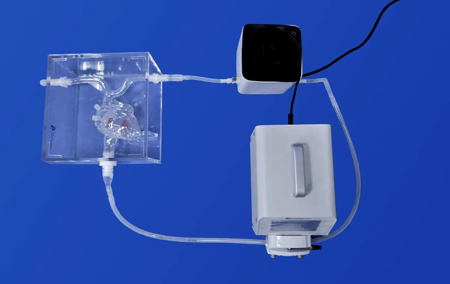
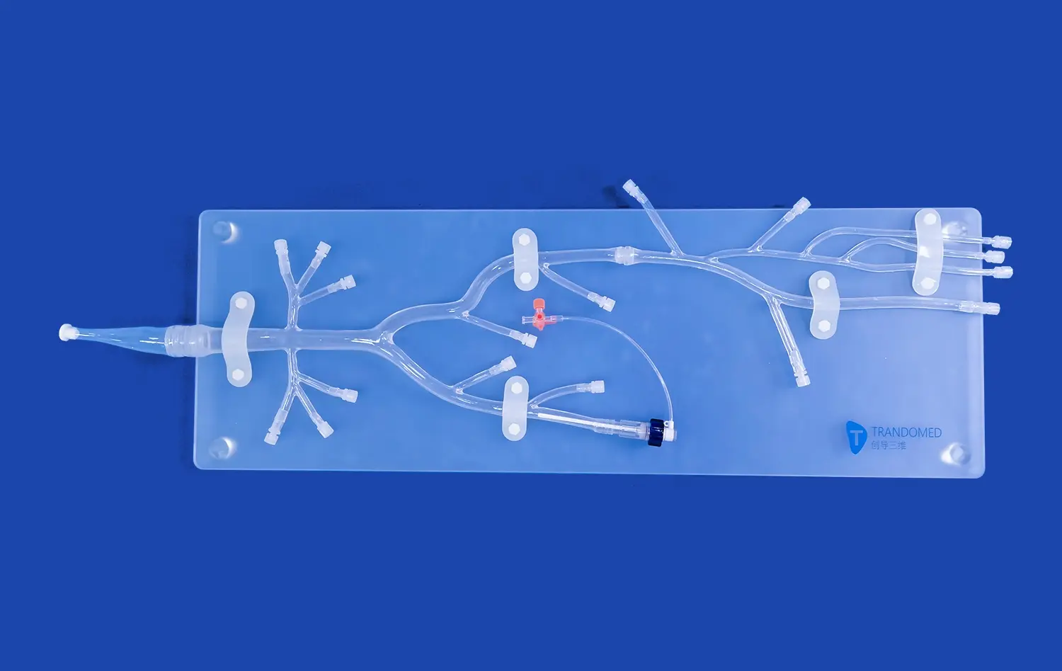
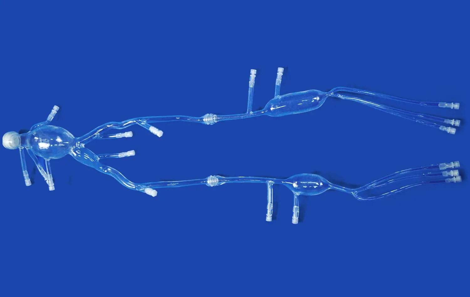
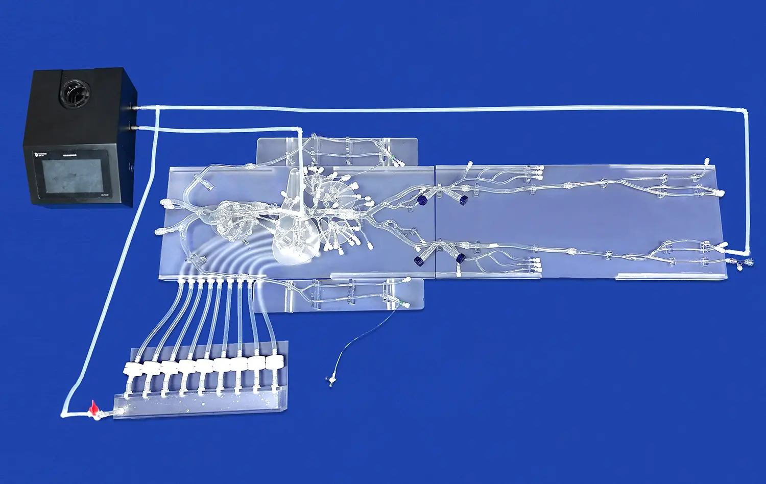
The Aortic Dissection Model (XXK004D) exemplifies this benefit by providing a comprehensive representation of major arteries involved in aortic dissection scenarios. Students can trace the path of blood flow from the ascending aorta through the aortic arch, down to the abdominal aorta and its branches, gaining a holistic understanding of the cardiovascular system's architecture.
Tactile Learning Experience
The tactile nature of physical models engages multiple senses, reinforcing learning through touch and sight. This multi-sensory approach is particularly beneficial for kinesthetic learners who grasp concepts more effectively through physical interaction. By allowing students to feel the texture and contours of the aortic wall, including the realistic depiction of the dissection lesion, these models create lasting mental imprints that enhance recall and understanding.
Comparative Analysis
Aortic dissection models facilitate comparative analysis between healthy and pathological states. Educators can use these models to demonstrate the stark differences between a normal aorta and one affected by dissection. This side-by-side comparison helps students identify key anatomical changes and understand how these alterations impact blood flow and overall cardiovascular function.
Teaching Dynamic Pathophysiology Through Simulation
Visualizing Disease Progression
Aortic dissection models serve as powerful tools for visualizing the progression of this potentially life-threatening condition. The realistic depiction of the dissection lesion in the thoracic aorta segment allows students to observe how the intimal tear propagates and affects the aortic wall layers. This visual representation helps learners grasp the concept of false lumen formation and understand how it compromises blood flow to vital organs.
Advanced models may incorporate features that simulate the dynamic nature of aortic dissection, such as adjustable flaps or color-coded sections representing areas of compromised blood flow. These interactive elements bring the pathophysiology to life, making complex concepts more accessible and memorable.
Understanding Hemodynamic Changes
Simulation using aortic dissection models enables students to explore the hemodynamic changes associated with this condition. By manipulating the model, learners can observe how the dissection affects blood flow patterns, pressure distribution, and perfusion to organs supplied by the affected aortic segments. This hands-on experience reinforces theoretical knowledge about the cardiovascular system's response to structural abnormalities.
Exploring Complications and Sequelae
High-fidelity aortic dissection models allow for the exploration of potential complications and sequelae associated with the condition. Educators can use these models to demonstrate how dissection can lead to aortic rupture, cardiac tamponade, or organ ischemia. By visualizing these critical scenarios, students develop a deeper appreciation for the urgency of accurate diagnosis and prompt intervention in aortic dissection cases.
Integrating Aortic Dissection Models into Medical Curricula
Enhancing Diagnostic Skills
Incorporating aortic dissection models into medical curricula significantly enhances students' diagnostic skills. These models provide a platform for practicing physical examination techniques, such as auscultation and palpation, in the context of aortic dissection. Students can learn to identify key clinical signs, such as pulse deficits or murmurs, associated with the condition.
Moreover, the models serve as excellent tools for teaching imaging interpretation. By correlating the physical model with various imaging modalities like CT angiography or MRI, students develop proficiency in recognizing radiological signs of aortic dissection. This integration of physical models with imaging studies bridges the gap between anatomical knowledge and clinical diagnosis.
Procedural Training and Skill Development
Aortic dissection models play a crucial role in procedural training for medical students and residents. These models provide a safe environment for practicing interventional techniques and surgical procedures related to aortic dissection management. Learners can rehearse critical skills such as:
- Endovascular stent graft placement
- Fenestration techniques
- Surgical repair of the aortic wall
The ability to practice these procedures on realistic models before encountering actual patients enhances confidence, improves technical skills, and ultimately contributes to better patient outcomes.
Interdisciplinary Learning
Aortic dissection models facilitate interdisciplinary learning by bringing together various medical specialties. These models can be used in collaborative educational sessions involving cardiology, vascular surgery, radiology, and emergency medicine teams. Such interdisciplinary approach mirrors real-world clinical scenarios, where the management of aortic dissection requires a coordinated effort from multiple specialties.
By engaging in simulations using these models, students and professionals from different disciplines can improve their communication skills, learn to work effectively in teams, and develop a holistic approach to patient care. This interdisciplinary exposure prepares future healthcare providers for the complex, collaborative nature of managing aortic dissections in clinical practice.
Conclusion
Aortic dissection models have revolutionized medical education by providing an immersive, hands-on learning experience that bridges the gap between theoretical knowledge and clinical practice. These models enhance anatomical understanding, facilitate the teaching of dynamic pathophysiology, and seamlessly integrate into medical curricula. By offering a safe environment for skill development and fostering interdisciplinary collaboration, aortic dissection models are instrumental in preparing the next generation of healthcare professionals to effectively diagnose and manage this critical cardiovascular condition. As medical education continues to evolve, the role of these advanced simulators in shaping competent, confident clinicians cannot be overstated.
Contact Us
For more information about our advanced Aortic Dissection Model (XXK004D) and other innovative medical simulation products, please contact Trandomed. Our team is dedicated to providing cutting-edge educational tools that enhance medical training and ultimately improve patient care. Reach out to us at jackson.chen@trandomed.com to explore how our customizable solutions can benefit your educational institution or healthcare facility.
_1736214519364.webp)
_1734507815464.webp)