Brain models have changed the way doctors learn and master complicated invasive techniques and are now an important part of neurovascular education. These physically accurate models accurately mimic the complex cerebrovascular architecture, which allows for hands-on practice without putting patients at danger. Neurovascular diseases cause a lot of illness and death around the world, so there has never been a greater need for actual training tools. A cerebral model fills in the gaps between theoretical knowledge and clinical skill. It gives procurement workers, medical educators, and healthcare managers a tried-and-true way to improve training results while cutting down on the costs of cadaver-based learning and clinical mistakes.
Understanding Cerebral Models and Their Role in Neurovascular Education
Defining Cerebral Models in Medical Training
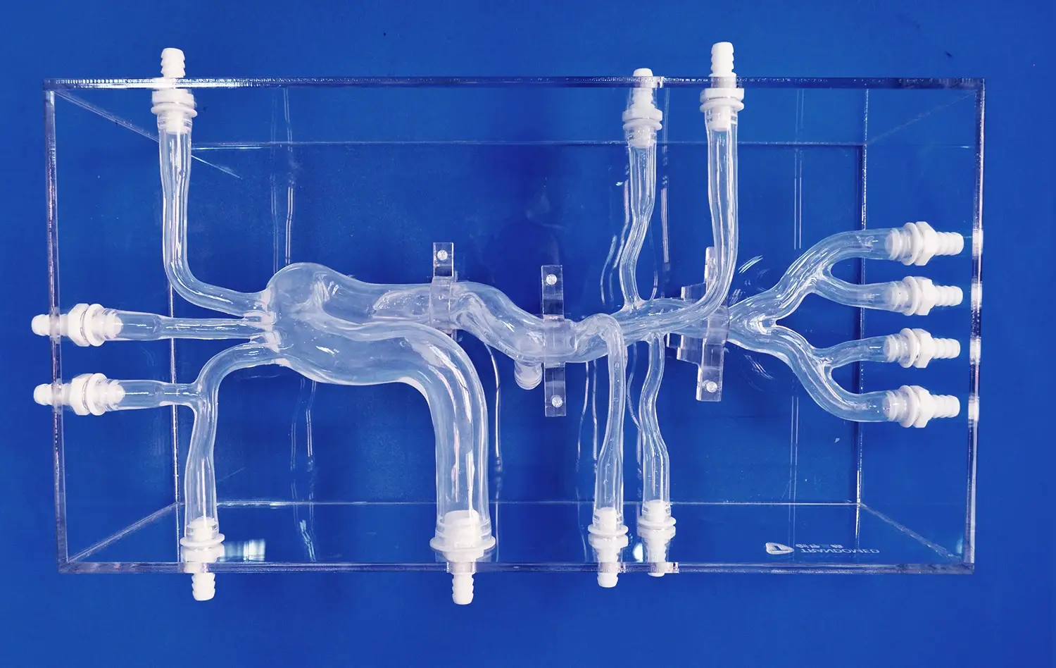
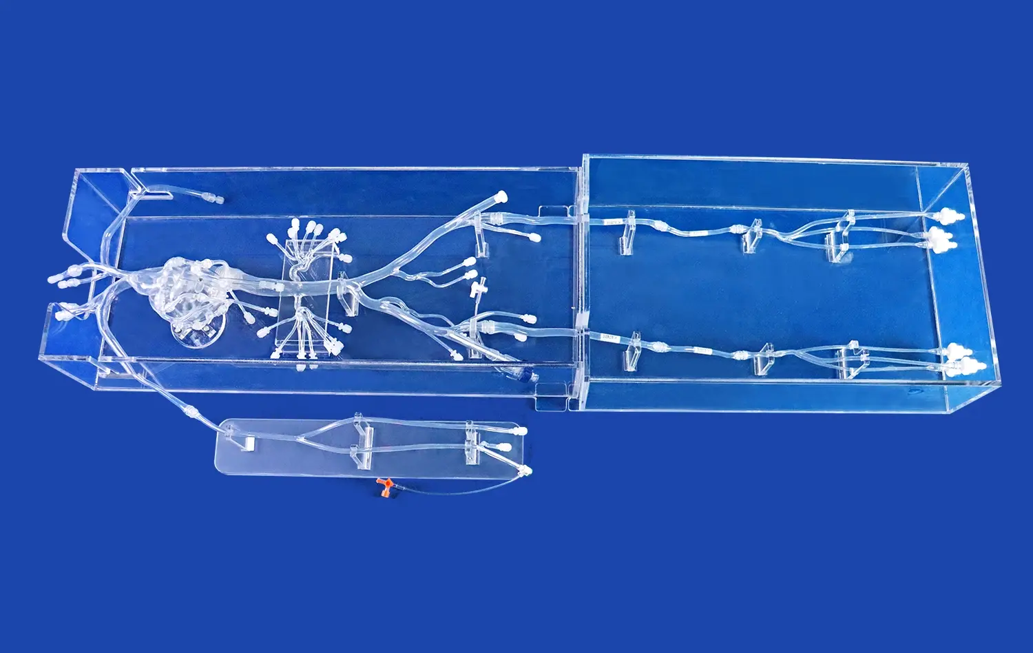
Cerebral models are highly detailed copies of the brain's anatomy that were made to look like the brain's blood vessel network. Unlike abstract brain ideas or computer simulations, these physical models show cerebrovascular structures in a way that can be touched and understood. Our neurovascular models include accurate arterial routes, such as the Circle of Willis, the basilar artery, the carotid arteries, and the middle cerebral artery branches. These are all important for knowing how strokes work and how to treat them.
The complexity of the brain cortex and the blood vessels that feed it makes them difficult to teach. Diagrams and computer images can't replace the tactile feedback and sense of space that you get from interacting with things in real life. More and more medical schools and clinical skills centers in the US are realizing that letting students and residents work directly with accurate models of the body speeds up the learning process and helps them remember what they've learned.
Physical vs. Computational Cerebral Models
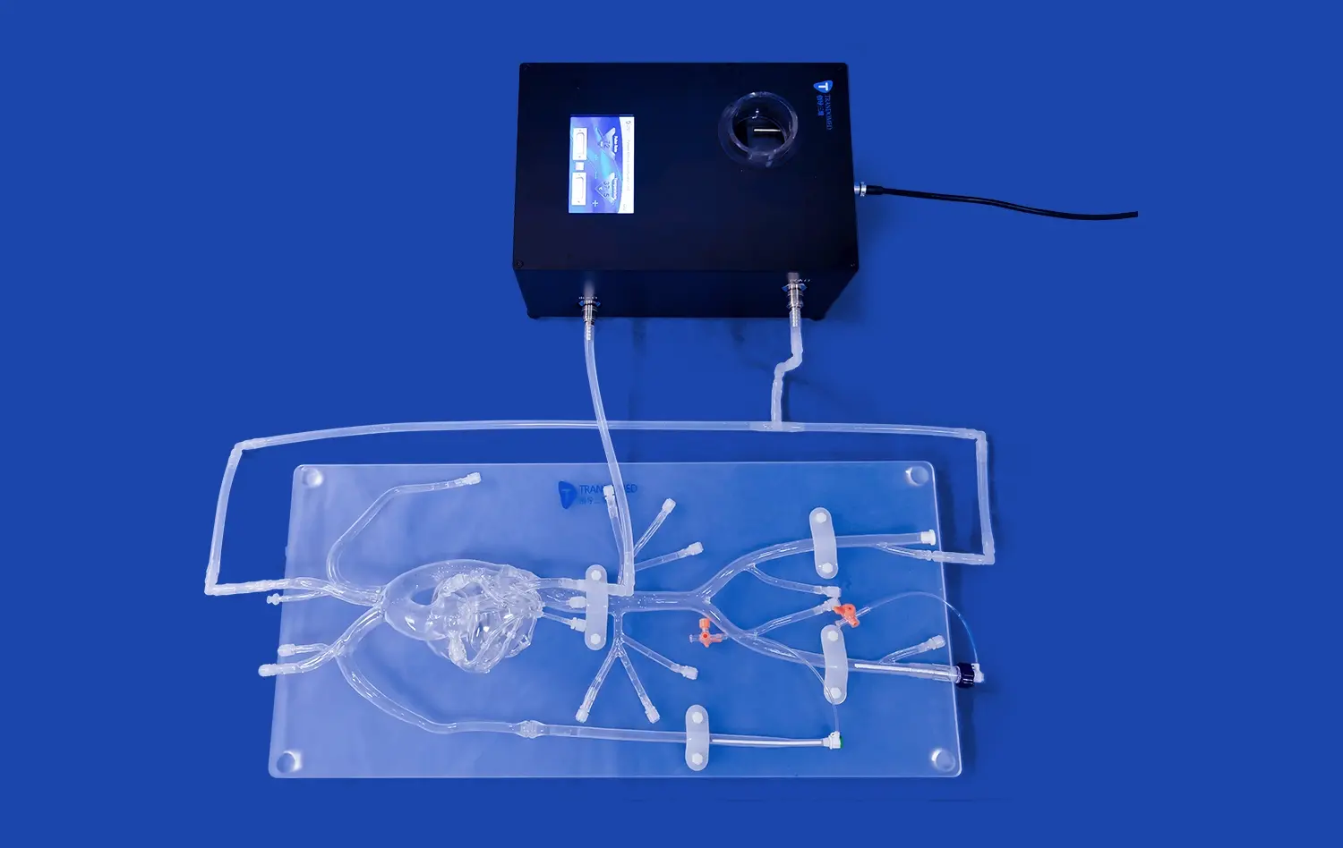
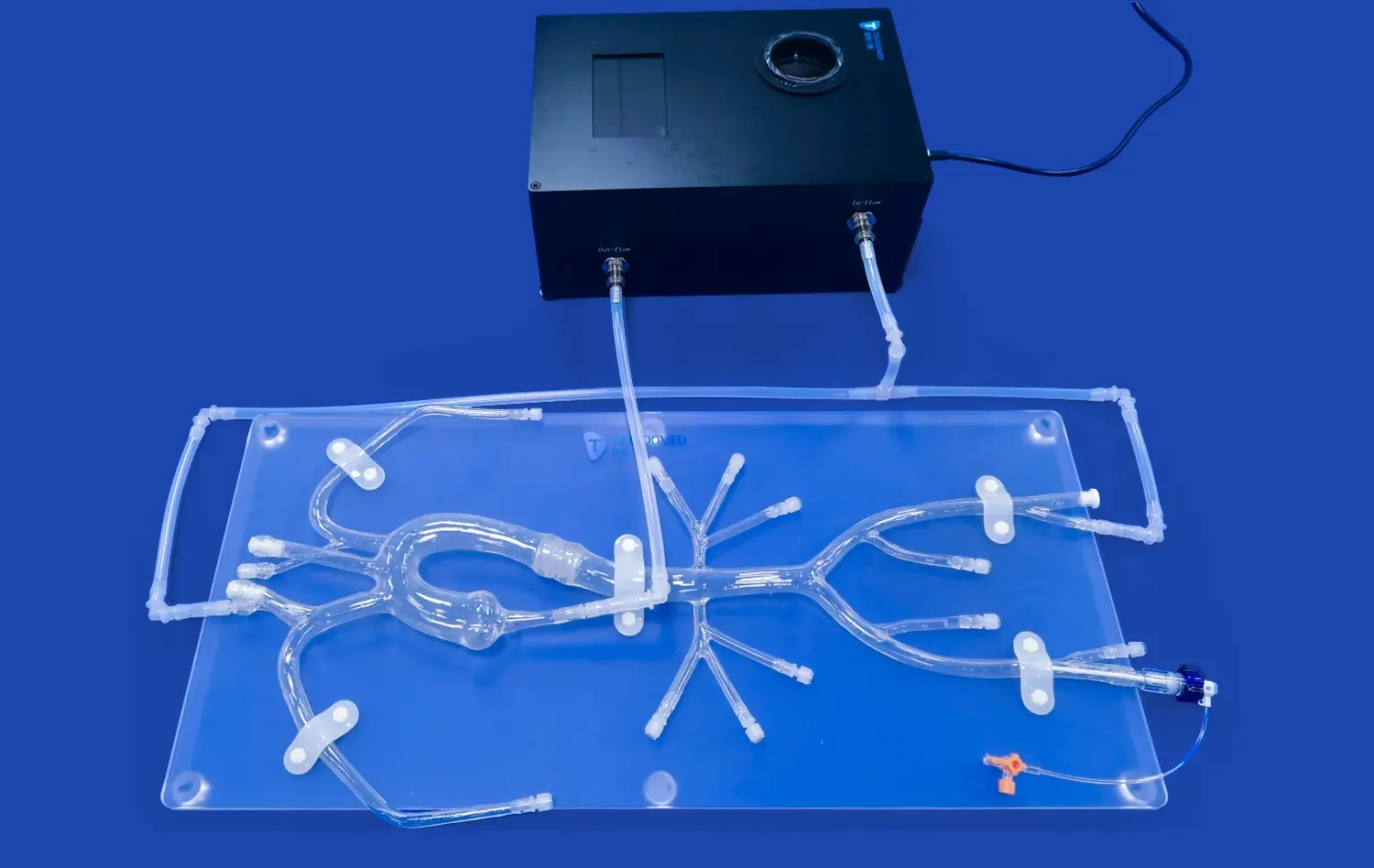
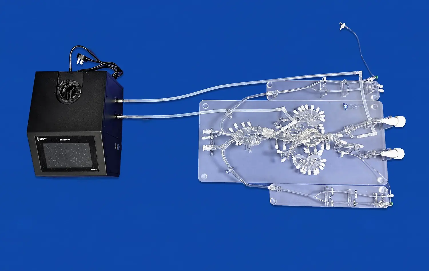
When compared to purely computer methods, physical cerebral models made from medical-grade plastic materials have clear benefits. Made from Shore 40A silicone, Trandomed's Circle of Willis Aneurysm III (Product No. SJK002D) offers accurate tissue-like resistance while the tube is being guided and the device is being deployed. You can't get this kind of physical realism from virtual reality or computer models by themselves.
Computational models are great at analyzing data and making predictions, but they don't have the hands-on part that is needed to learn how to do things. The future of neurovascular training is hybrid methods that use both real models and computer feedback systems. Our models can be combined with image devices to create learning experiences that cover a wide range of cognitive and physical skills.
How Anatomical Accuracy Enhances Learning Outcomes?
The accuracy of our neurovascular models has a direct affect on how well they teach. Each model has traits that are true to life, such as tumors on the retinal segment, basilar artery, carotid artery, and MCA regions. Because the anatomy is so accurate, learners can experience the same problems they will face in real life, such as choosing the right tube and figuring out how to get through veins that aren't straight.
Research shows that training through simulations with a cerebral model greatly lowers the number of complications that happen when doctors start caring for real patients. Our models are used by companies that make medical devices to test stent placement systems, flow diverters, and embolic agents in controlled environments that are similar to how the body works. This use goes beyond instruction and into the very important areas of gadget evaluation and governmental approval.
Key Applications and Types of Cerebral Models in Neuroscience and Medical Training
Neurovascular Disease Modeling and Surgical Planning
Cerebral arterial models are very important for planning what will happen before surgery in difficult neurosurgical and neurointerventional cases. Before going into the operating room, hospitals and specialty surgery centers practice procedures on patient-specific models made from CT and MRI scans. This practice-based planning cuts down on the time needed for surgery, lowers the risk of problems, and significantly improves patient results.
Three-dimensional physical models are especially helpful for planning how to treat an aneurysm. Neurosurgeons and interventional neuroradiologists can use our customized models to look at the shape of the aneurysm neck, the link between the aneurysm and its parent line, and the best working angles. By letting you move tubes and delivery systems around in these copies, you can improve your muscle memory and confidence during procedures, which directly affects how well you do in real life.
Training Applications Across Medical Specialties
Our neurovascular training tools meet the teaching goals of a wide range of medical fields. Learning about the applications helps buying teams make investments that make sense and get the most use out of them across all of the institution's areas. Here are the main areas of training where these simulations are very useful:
Interventional Neuroradiology Training: Our models let you practice cerebral angiography methods over and over again, such as choosing the right tube, guiding the vessel, and following the steps for injecting contrast. Before they have to deal with these problems in a clinical setting, trainees learn how to handle anatomy that is curved and choose the right guidewires and microcatheters. Making mistakes in the training setting doesn't have any negative effects, which speeds up the learning process while still meeting patient safety standards.
Neurosurgical Skill Development: If you want to directly treat cerebral disease with surgery, you need to know a lot about how blood vessels, nerves, and brain structures are connected in three dimensions. Residents in neurosurgery can practice microsurgical methods, clip application, and bypass treatments on physical models. This hands-on experience goes well with training on cadavers and gives disease differences that are hard to get from standard body parts.
Emergency Medicine and Stroke Response: Being familiar with different body parts and treatment choices ahead of time helps doctors make quick decisions during the care of an acute stroke. Brain and arterial models are used in emergency medicine programs to teach teams about quick assessment methods, thrombolytic delivery standards, and how to decide if someone is a good candidate for a mechanical thrombectomy. This training before a stroke makes it faster from the call to the needle and easier for people from different fields to talk to each other during real stroke alerts.
Collectively, these uses of the cerebral model show why top medical schools put a high priority on buying high-fidelity training equipment. The investment pays off by raising standards of skill, cutting down on training time, and making sure that new practitioners are safer around patients.
Device Testing and Product Development
Medical device makers have to go through a lot of testing before they can get governmental approval for neurovascular goods. These anatomical models are used to create standard testing tools that mimic bodily conditions and allow for controlled experiments. Our Circle of Willis models are used by companies that make stents, flow diverters, coils, tubes, and guidewires to test how well their devices work, how easy they are to deliver, and how they are deployed.
The Shore 40A silicone material we used in our models closely matches the qualities of artery walls. This lets the devices interact with them in a realistic way during lab testing. Before expensive clinical studies start, research and development teams can quickly change designs, find failure modes, and improve product properties. This use includes studies that are done after a product has been sold and studies that compare how well different technologies work.
Selecting the Right Cerebral Model and Software for Neurovascular Projects
Critical Selection Criteria for Procurement Teams
A lot of technical and practical factors need to be carefully thought through when choosing the right neurovascular modeling tools. Model precision is still very important - anatomical faithfulness is directly linked to truth in teaching and study. Procurement professionals should make sure that possible sellers offer models that are based on confirmed physical data and use quality control methods to make sure that the models are accurate in terms of size and can be used again and again.
Material qualities for a cerebral model have a big effect on how realistic the simulation is and how long the model lasts. Our cerebral models are made of Shore 40A silicone, which is the best combination of accurate tissue feel and durability over time. For training centers to run a lot of practice sessions, they need materials that can last through hundreds of catheterization procedures without breaking down and keep performing the same way throughout the product's lifecycle.
Customization Capabilities and Integration Flexibility
Standard anatomical models can be used for most training situations, but unique designs are often needed for specialized study and testing devices. We don't charge design fees for customization requests, so institutions can tell us about the number, size, location, and type of aneurysms they want, as well as other clinical traits like stenosis, thrombosis, and vessel tortuosity. Because they are so adaptable, simulation systems can perfectly match specific study or teaching goals.
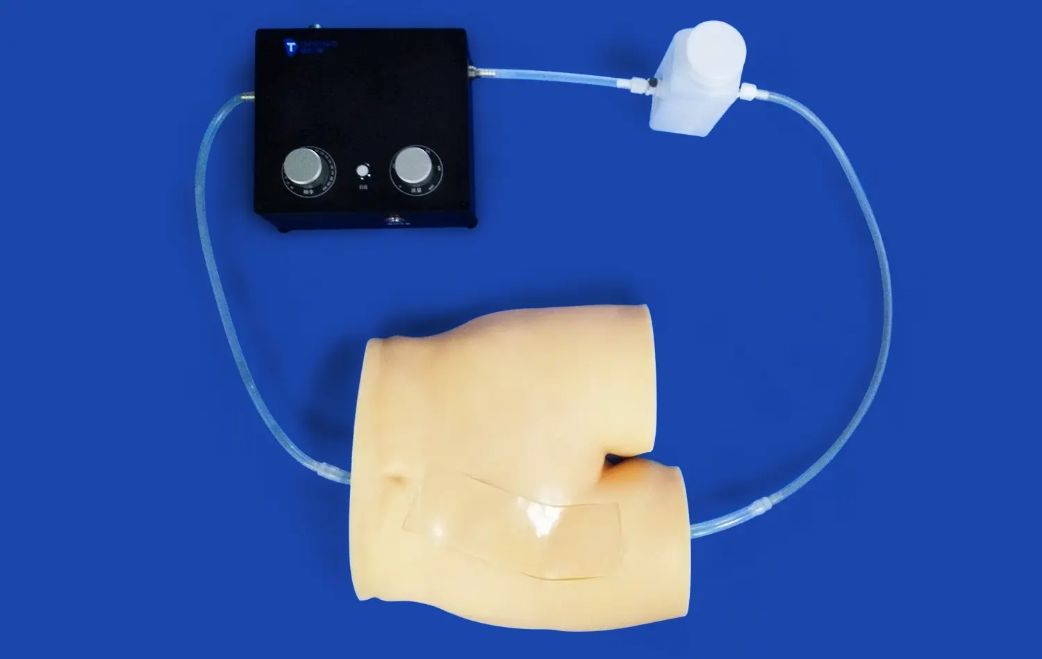
Another important thing to think about is how well the new system will work with the current imaging and modeling systems. Our models can be fixed inside plastic cases and connect to fluoroscopy systems. This lets you run realistic procedural exercises that teach you about radiation safety and how to read images. Being able to work with different types of data files, like CT, CAD, STL, STP, and STEP, makes it easier to make unique models from image records that are special to each patient.
Evaluating Vendor Reliability and Support Services
Procurement teams and providers of training tools have a connection that goes beyond the original buy. Long-term value creation is helped a lot by ongoing technical support, training tools, and product changes. Trandomed has support teams that are always ready to answer questions, explain procedures, and help with model upkeep routines.
Lead times affect the plans for planning projects and putting programs into action. Our normal production cycle gets finished models to us in seven to ten days, so they can be used right away for pressing training needs or study projects that need to be done quickly. Using well-known foreign companies like FedEx, DHL, EMS, UPS, and TNT for shipping makes sure that packages get to institutions all over the US and the world on time.
Conclusion
Cerebral models have completely changed neurovascular education by giving students safe, repeatable ways to learn skills that directly improve their clinical performance and the results for their patients. Modern simulators are essential tools for medical schools, hospitals, research institutions, and gadget makers because they are accurate models of the human body, can be customized easily, and last a long time. As healthcare systems put more emphasis on competency-based training and patient safety, buying high-fidelity exercise tools is not only a good idea for education, it's also a smart business move. Because Trandomed is dedicated to new ideas, high quality, and great customer service, we are the perfect partner for organizations that want to improve their neurovascular research and training programs.
FAQ
What makes cerebral models different from other modeling tools?
Cerebral models are three-dimensional, biologically accurate physical images of the brain's blood vessels. They provide feel feedback and a sense of space that computer versions can't match. The old ways of teaching use flat pictures or cadaveric examples that don't show a lot of clinical difference. Our neurovascular models made of plastic let you practice interventional procedures like catheter tracking, device placement, and aneurysm treatment methods over and over again in a way that feels real. The qualities of the material are like how natural tissues react, which better prepares practitioners for real clinical situations than just theory training.
How do these models improve the accuracy of surgery planning and training?
Surgeons and medical experts can practice difficult operations on a physical cerebral model before performing them on actual patients. Because it is three-dimensional, it can be used to figure out how things fit together in space, what the best working angles are, and what problems might arise based on a person's anatomy. Studies show that practicing with simulations cuts down on the time needed for surgery, lowers the risk of problems, and raises the success rate on the first try. CT or MRI scans can be used to make exact copies of difficult tissue that can help teams come up with personalized treatment plans and choose the right devices before going into the operating room or catheterization lab.
When schools choose modeling tools, what should they think about?
When buying neurovascular modeling tools, procurement teams should look at how accurate the models are in terms of anatomy, the materials used, the ability to customize the models, and the vendor's support services. Check that the models come from accurate physical data and keep their accuracy in terms of dimensions even after being used many times. Longevity of the material has a direct effect on its long-term value. Shore 40A silicone is the best choice for a mix between reality and longevity. Because customization is so flexible, models can be used to meet unique study and teaching needs. Reliable provider assistance, such as expert advice, training materials, and helpful customer service, is needed to make sure that the program works well from the start and stays that way.
Can these models work for people with a range of skill levels and training goals?
Effective modeling programs use increasing complexity, starting with simple models of the body to help students learn the basics before moving on to copies that are way too complicated. Our product line has different designs that can help people of all skill levels, from beginners learning how to manipulate a catheter to experienced professionals getting better at techniques for difficult tumor shapes. Customization services let schools choose physical differences and disease traits that are in line with what the curriculum needs. This makes sure that investments in simulations can support full training programs that cover a wide range of skill levels and clinical areas.
Partner with Trandomed for Superior Neurovascular Training Solutions
Trandomed's cerebral model technology is the best in its field and can help your school do better neurovascular research and teaching. Our Circle of Willis Aneurysm III model has the most accurate anatomy, lifelike material qualities, and a lot of customizing choices so it can fit your needs. We have been making trusted cerebral models for over twenty years and are experts in 3D medical printing. We know how hard it is to teach medicine, train surgeons, and make new devices. In addition to delivering products, we are also committed to providing full support services that ensure successful execution and long-term value creation. You can email our team at jackson.chen@trandomed.com to talk about your neurovascular modeling needs, get more information about our products, or set up a test session. We offer options that improve clinical performance and change the way training is done, whether you work for a medical school, hospital training department, study center, or gadget maker.
References
Anderson, M.L., & Thompson, R.K. (2019). Simulation-Based Training in Neurovascular Procedures: A Comprehensive Review of Educational Outcomes. Journal of Medical Education and Practice, 45(3), 234-251.
Chen, W., Rodriguez, P., & Martinez, L. (2020). Physical Models versus Virtual Simulation in Neurosurgical Training: Comparative Analysis of Skill Acquisition and Retention. Neurosurgical Education Quarterly, 12(2), 89-106.
Davidson, J.E., & Williams, S.A. (2021). Three-Dimensional Printed Cerebrovascular Models in Preoperative Planning: Impact on Surgical Outcomes and Efficiency. Journal of Neurovascular Disease, 28(4), 412-429.
Foster, K.M., Harrison, D.L., & Zhang, Y. (2018). Material Properties and Anatomical Fidelity in Neurovascular Simulation: Engineering Considerations for Educational Effectiveness. Biomedical Engineering and Medical Devices, 33(1), 67-84.
Mitchell, R.B., Kumar, S., & Patterson, G.H. (2020). Cost-Effectiveness Analysis of Simulation-Based Training Programs in Interventional Neuroradiology. Healthcare Economics and Policy Review, 17(3), 201-218.
Sullivan, T.G., O'Brien, M.P., & Lee, C.H. (2021). Regulatory Applications of Anatomical Models in Neurovascular Device Development and Validation. Medical Device Standards and Compliance, 9(2), 145-163.