Why Electrophysiology Simulation Matters in Cardiology Training?
2026-05-27 09:00:03
The way we train the next crop of cardiologists has changed a lot because of electrophysiology simulation. Even though traditional methods are useful, they often put people at risk while they are learning. This problem can be solved with advanced modeling tools, especially the venous cardiac electrophysiology model, which very accurately models the heart's electrical system. These models make it possible for doctors to learn how to use catheters, do mapping, and do ablation treatments in a safe setting. A big change in medical education is being able to practice difficult interventions over and over again without putting patient safety at risk. This prepares doctors to handle real-life situations with confidence and accuracy.
Understanding Venous Cardiac Electrophysiology Models in Cardiology Training
What Makes Venous Models Different from Other Cardiac Simulations?
The cardiac venous system is very important for electrophysiology treatments, but in many training programs, it doesn't get as much attention as arterial pathways. The inferior vena cava, superior vena cava, right atrium, right ventricle, and subclavian vein are just a few of the structures that venous models accurately copy. These are important parts for catheter-based treatments. These models are different from arterial simulations because they focus on the difficulties of working with veins during pacemaker implantation, cardiac resynchronization therapy, and diagnostic EP tests.
Key Anatomical Features Replicated in Modern Simulators
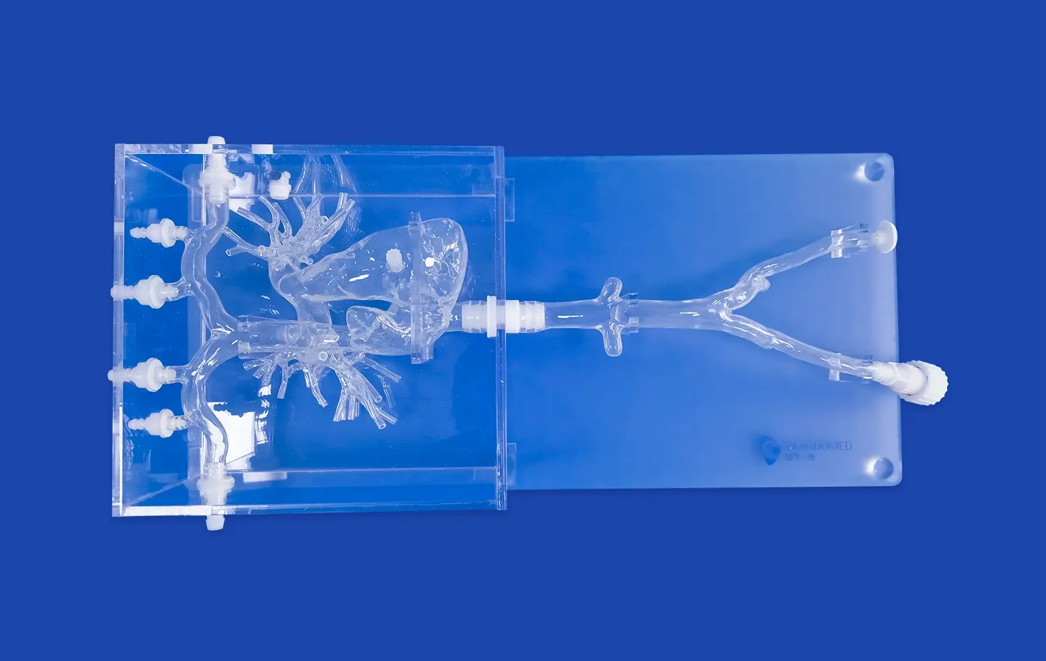
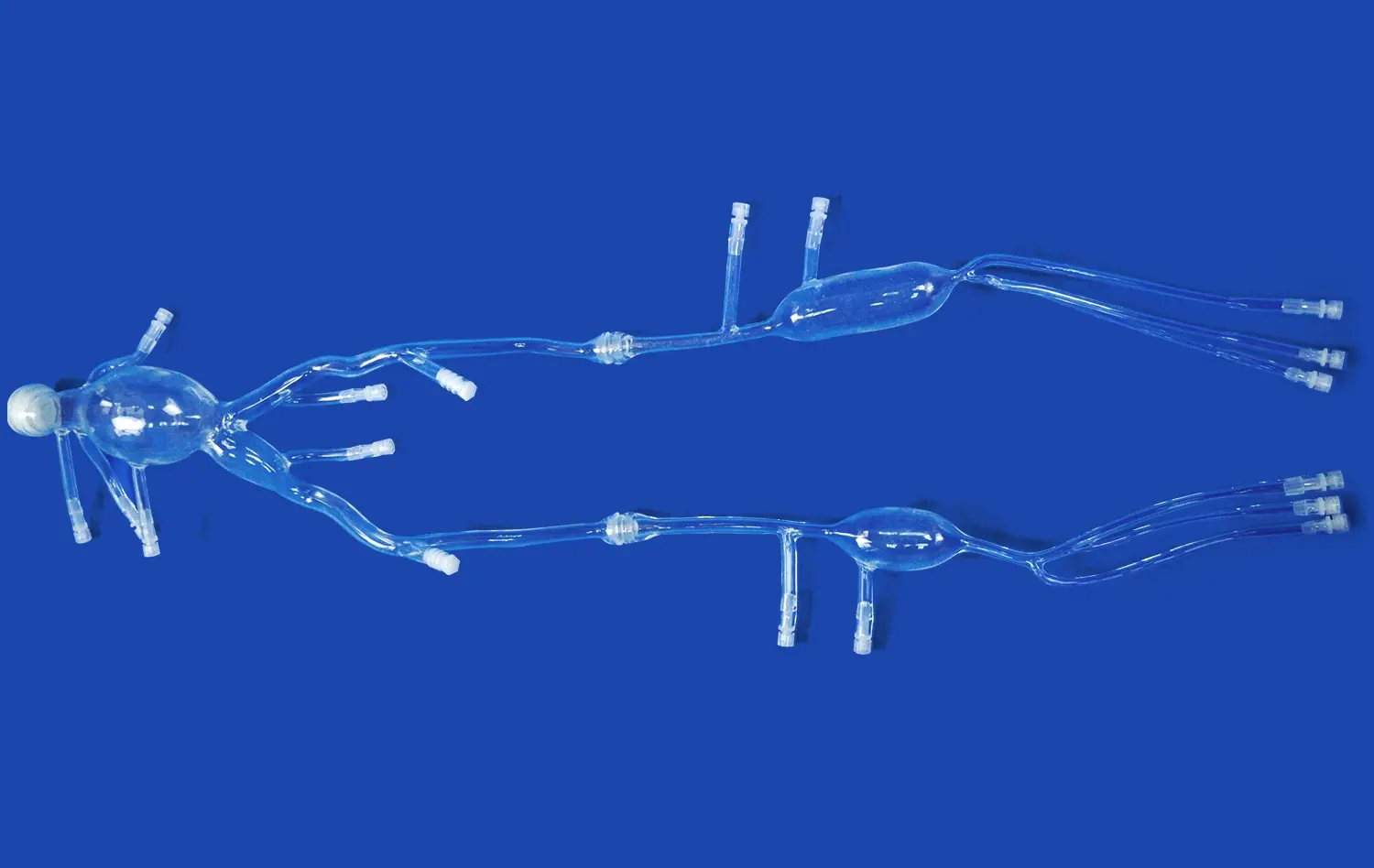
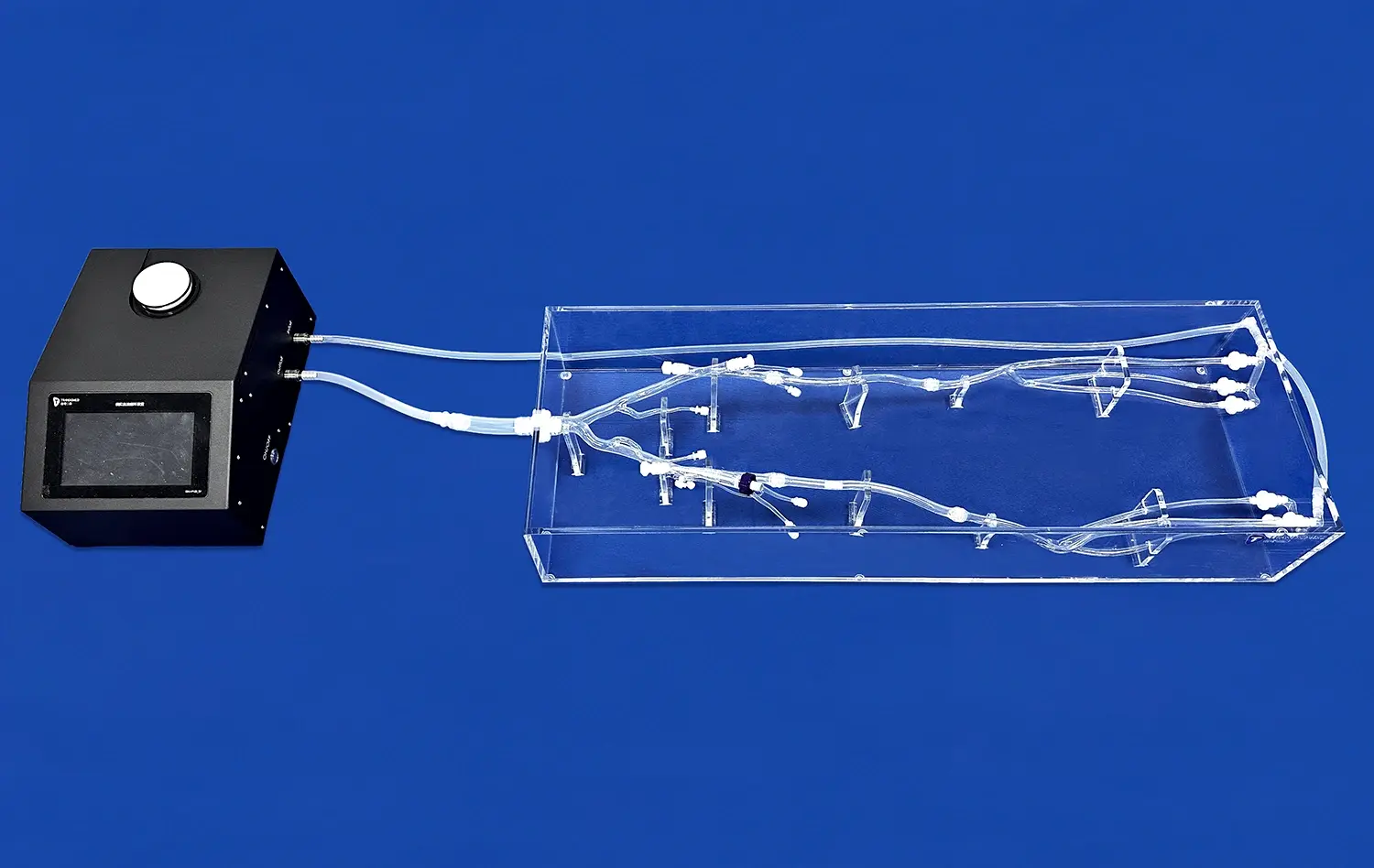
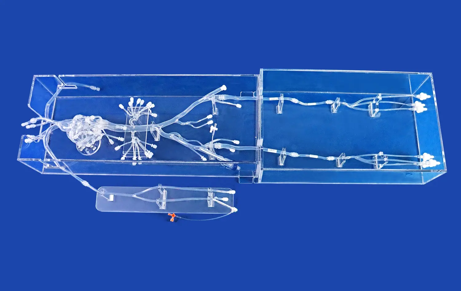
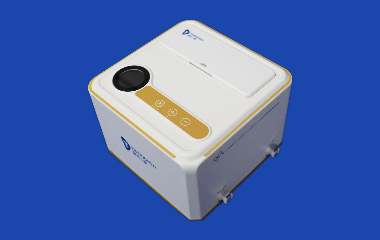
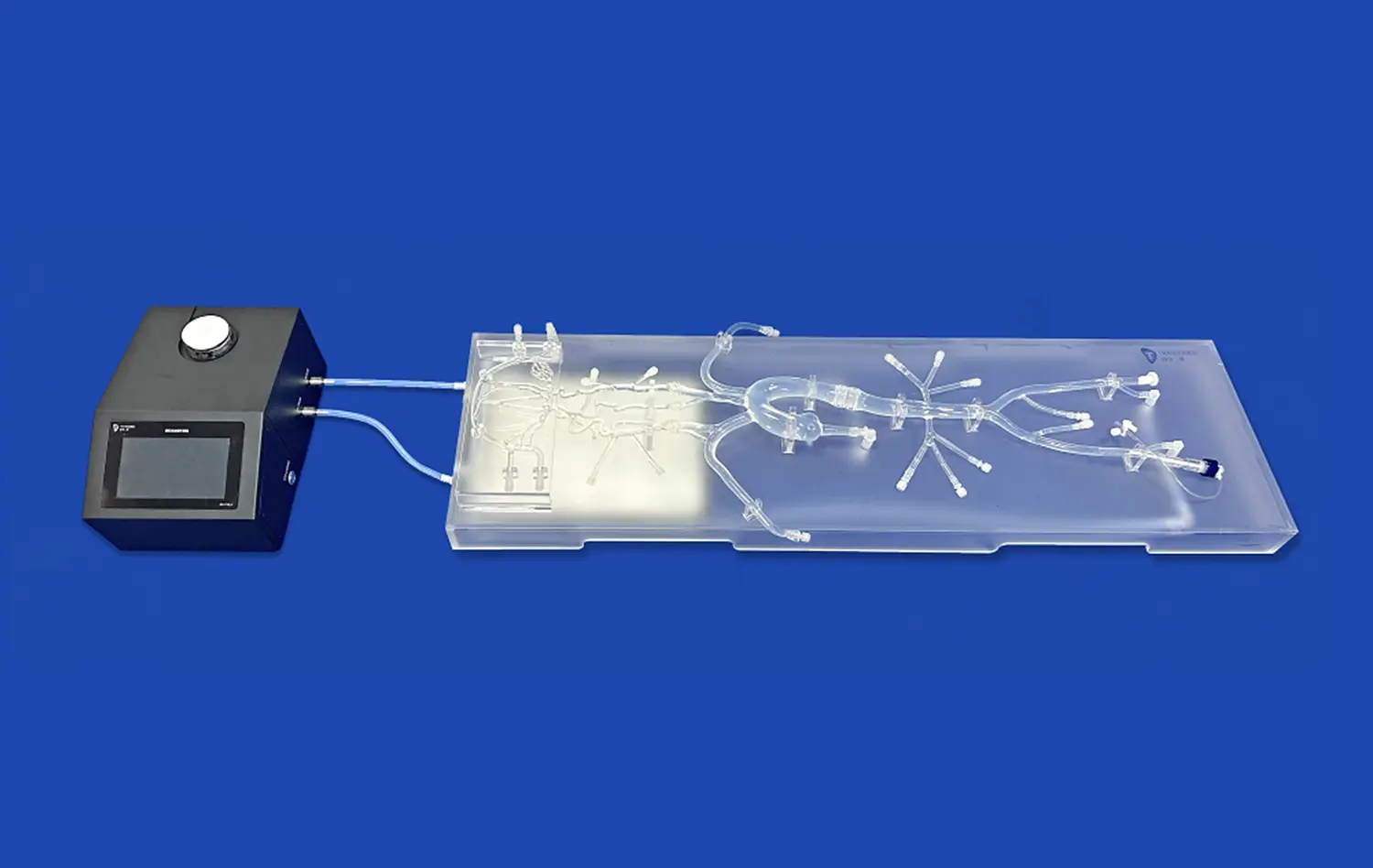
Thanks to progress in simulation technology, it is now possible to record intricate anatomical features that were not possible before. Trandomed's Venous Cardiac Electrophysiology Model (XXS004) is a good example of this change. It is made of silicone and has a Shore 40A durometer that makes it respond like human tissue. This model has all the important vein structures used in electrophysiology treatments, and it gives you feedback that feels a lot like the anatomy of a real patient. The choice of material makes sure that it will last through multiple training classes while still having realistic handling characteristics that help train people for clinical practice.
How Simulation Technology Enables Risk-Free Practice
Simulations make a place to learn where mistakes are seen as opportunities to learn rather than threats to patient safety. Trainees can work on manipulating catheters, learning to spot anatomical landmarks, and building muscle memory for more difficult treatments. With this technology, teachers can gradually add more difficult situations, ranging from simple changes in structure to difficult pathological conditions, helping students get better over time. Modern simulators are flexible enough that training scenarios can be changed to meet specific learning goals, such as teaching medical students basic ideas or improving advanced methods for doctors who have been practicing for a while.
Advantages of Electrophysiology Simulation Over Traditional Training Methods
Addressing the Limitations of Traditional Apprenticeship Models
In traditional cardiology training, you learn a lot by watching processes and then taking on more responsibility while being supervised. This apprenticeship method has some good points, but it depends on how many patient cases are available and raises safety issues while skills are being learned. Trainees may have to wait months before they see certain arrhythmias or differences in anatomy, which makes learning less reliable. Simulation gets rid of these problems by giving practitioners instant access to a wide range of situations, which lets them get better before they work with real people.
Creating Repeatable and Standardized Learning Experiences
One big benefit of modeling is that it can be used over and over again. The same anatomical situation can be practiced over and over, which helps students improve their skills in a planned way. This kind of repetition builds trust and skill in ways that one-time clinical exposure can't. Standardization also helps with evaluations because teachers can use the same factors and situations to grade all of their students. Training programs can set standards for measurable competency that make sure graduates meet consistent quality standards before they do processes on their own.
Measurable Improvements in Skill Acquisition and Retention
Major cardiac training centers have done research that shows that practitioners who are trained in simulations become proficient in procedures faster than those who only use traditional methods. Studies have shown that the number of complications is lower when newly trained doctors go from simulators to real patients. Being able to make mistakes safely, get feedback right away, and fix your skill speeds up the learning process by a lot. Also, retention rates go up because students are involved in real-life situations that make better mental connections than just learning theory.
These changes to training are very helpful for companies that make medical devices. When making new EP catheters, ablation systems, or mapping technologies, it is very helpful to test them on accurate simulation models. Simulations allow companies to show potential customers how a gadget works before it goes through clinical trials. This speeds up the market entry and adoption process. Because simulator testing can be repeated, it also helps with regulatory submissions by giving uniform performance data across controlled conditions.
Selecting the Right Venous Cardiac Electrophysiology Model for Your Organization
Evaluating Accuracy and Anatomical Fidelity Requirements
Not every computer venous cardiac electrophysiology model is as realistic or useful as the others. Companies need to figure out what amount of anatomical detail is needed for the training they want to give. For teaching basic anatomy, simpler models may be enough, but for teaching advanced catheter handling, exact copies with the right tissue properties are needed. The venous cardiac electrophysiology model should not only show the anatomy as it appears to the naked eye, but it should also show how the doctors feel when they are doing procedures. Trandomed meets this need with technology that is based on a lot of human CT and MRI data and uses reverse 3D modeling to show the body accurately.
Compatibility with Existing Training Infrastructure and Workflows
Problems with integration can make even the best modeling technology less useful. Teams in charge of buying things should think about how new models will fit in with current training programs, physical areas, and equipment stocks. Some simulators need special imaging or recording gear to work, while others can be used on their own as teaching tools. The XXS004 model's design makes it easy to use in a variety of teaching settings, from medical schools to demonstration labs for device manufacturers. Because it can be used over and over, it's good for high-volume training programs that don't need to change it often.
Customization Capabilities and Service Support Options
Different groups and uses have very different training needs. Some research labs need anatomical differences that show certain diseases, while medical schools might prefer normal anatomy with common variations. Trandomed gives a wide range of customization options and can work with data files in CT, CAD, STL, STP, and STEP formats to make models that exactly match your needs. A big problem that makes it harder to get unique solutions is that the company doesn't charge for design changes. Short production times (usually seven to ten days) make sure that training plans can be put in place without any long delays.
Procurement considerations extend beyond initial acquisition to ongoing support relationships. Companies should check how responsive their vendors are, how easy it is to get technical help, and keep their rules up to date. Training program investments are safe when they work with makers who understand the problems in medical education and are committed to making products better over time. With 20 years of experience in medical 3D printing technology, Trandomed is able to offer not only goods but also information on new ways to teach and technology that is getting better.
Overcoming Challenges and Ensuring Effective Model Implementation
Balancing Realism with Practical Training Constraints
It is always hard to get perfect anatomical recreation while keeping the system usable. Too many details in a model can make it break easily or be too expensive for most people to afford, and too few details in a model doesn't help teach people properly. Organizations need to find the best balance point where models are realistic enough to help people apply what they've learned in the real world without adding too much complexity or cost. Silicone materials, like the ones used in the XXS004, are a great compromise because they allow for realistic tissue contact and last for hundreds of practice sessions.
Integrating Simulation into Comprehensive Curriculum Design
Buying simulation equipment does not ensure that the training program will work. For implementation to work well, the program needs to be carefully planned so that simulations work with other teaching methods instead of replacing them. Programs should figure out which skills simulations help with the most and then create organized learning progressions for those skills. Simulations work best when combined with classroom teaching, clinical observation, and supervised patient care. This creates a well-rounded learning experience that improves knowledge, skills, and judgment all at the same time.
Maintaining Relevance Through Continuous Updates and Evolution
Clinical methods and medical technology are always changing, so training tools need to keep up. Organizations should set up ways to check the usefulness of computer venous cardiac electrophysiology models on a regular basis and update hardware or software as needed. Having relationships with vendors that allow for continuing customization and modification makes training investments last longer. This method is shown by Trandomed's ability to reconstruct atrium segments using real human CT data. This lets organizations change their training models when new information about anatomy or procedures comes out.
Another thing to think about when implementing is technical proof. Training programs should set up procedures to make sure that the performance of simulators correctly mirrors what happens in the real world. For this, a skilled practitioner might look it over and see if it matches up with clinical outcome data, or formal studies could be used to see how well skills learned in modeling are applied in real life patient care. Validation that can be proven in writing makes a program more trustworthy with accreditation bodies, school leaders, and trainees.
Maximizing ROI with Electrophysiology Simulation in Cardiology Training
Quantifying Training Efficiency Gains and Cost Savings
Simulation investments deliver returns through multiple channels. Reduced training time translates directly to cost savings as practitioners reach competency faster. Decreased complication rates among newly trained physicians avoid expensive adverse event management and liability exposure. Improved first-case success rates reduce procedure times and resource utilization. Organizations should track these metrics systematically to document simulation program value and justify continued investment.
Research institutions realize returns through enhanced experimental capabilities. Biomedical studies investigating arrhythmia mechanisms, testing pharmacological interventions, or validating new device concepts benefit from standardized, reproducible simulation platforms. The ability to conduct multiple experimental iterations quickly accelerates research timelines and publication productivity. Device manufacturers similarly achieve faster development cycles when prototype testing occurs on validated simulation models before expensive clinical trials.
Strategic Procurement and Vendor Relationship Management
Maximizing value requires strategic procurement approaches beyond simple price comparison. Organizations should evaluate total ownership costs including maintenance, consumables, training support, and customization services. Trial opportunities allow hands-on assessment before commitment, reducing implementation risk. Trandomed accommodates this need through flexible engagement models that let organizations experience simulation technology in their specific training contexts before making purchasing decisions.
Payment terms and delivery logistics affect implementation success. Understanding lead times helps organizations plan training program launches realistically. The XXS004 model's seven-to-ten-day production schedule supports relatively rapid deployment compared to custom solutions requiring months of development. Multiple shipping options through major international carriers provide flexibility for urgent needs or coordinated multi-site implementations.
Long-term vendor partnerships create opportunities for continuous improvement and innovation access. Manufacturers like Trandomed, with extensive experience developing medical simulators, bring valuable insights to training program design. Their understanding of emerging technologies, material science advances, and pedagogical research can inform organizational strategy beyond individual product transactions. These relationships transform vendors into collaborative partners invested in client success rather than mere equipment suppliers.
Conclusion
Electrophysiology simulation represents essential infrastructure for modern cardiology training, addressing fundamental limitations of traditional apprenticeship models while enhancing safety, efficiency, and skill development. The venous cardiac electrophysiology model specifically fills a critical gap in preparing practitioners for catheter-based procedures involving cardiac venous anatomy. Organizations that invest strategically in high-fidelity simulation technology position themselves at the forefront of medical education and research. Success requires careful model selection aligned with specific training objectives, thoughtful curriculum integration, and ongoing commitment to validation and improvement. The returns on these investments manifest through accelerated competency development, reduced clinical risks, and enhanced research capabilities—benefits that ultimately improve patient care and advance cardiovascular medicine.
FAQ
What procedures can be practiced using a venous cardiac electrophysiology model?
These specialized models support training in catheter navigation through venous pathways, electrophysiological mapping of cardiac electrical activity, pacemaker lead placement, cardiac resynchronization therapy device implantation, and catheter ablation techniques. The anatomical accuracy of quality models allows practitioners to develop skills applicable directly to patient care, including recognizing anatomical landmarks, manipulating catheters safely, and understanding spatial relationships between venous structures and cardiac chambers.
How does the bidomain model relate to cardiac electrophysiology simulation?
The bidomain model represents a mathematical framework describing cardiac tissue electrical properties, serving as the computational foundation for many electrophysiology simulations. This model treats cardiac tissue as two interconnected domains—intracellular and extracellular spaces—each with distinct electrical properties. While primarily used in computational simulations, bidomain model principles inform the design of physical simulation models by defining realistic electrical behavior that training devices should replicate during procedure practice.
What distinguishes an electrophysiology study from standard ECG testing?
An electrocardiogram provides a surface recording of cardiac electrical activity, offering overview information about heart rhythm and conduction. An electrophysiology study involves invasive catheter placement within the heart to record electrical signals directly from cardiac tissue. This invasive approach delivers detailed information about conduction pathways, arrhythmia mechanisms, and specific treatment targets unavailable through surface ECG. EP studies require specialized procedural skills that simulation models help practitioners develop before performing invasive testing on patients.
Partner with Trandomed for Advanced Cardiac Simulation Solutions
Trandomed combines two decades of medical 3D printing innovation with deep understanding of cardiology training needs. Our venous cardiac electrophysiology model manufacturer capabilities deliver custom solutions without design cost penalties, accelerating your program implementation. The XXS004 model features anatomically precise silicone construction, rapid production timelines, and extensive customization options based on your CT data specifications. We invite procurement managers, training directors, and research coordinators to explore how our simulation technology enhances learning outcomes and operational efficiency. Contact Jackson Chen at jackson.chen@trandomed.com to discuss your specific requirements and arrange product evaluation.
References
Zipes, D.P., Jalife, J. (2021). Cardiac Electrophysiology: From Cell to Bedside (8th ed.). Philadelphia: Elsevier.
Josephson, M.E. (2020). Clinical Cardiac Electrophysiology: Techniques and Interpretations (5th ed.). Philadelphia: Wolters Kluwer.
Anderson, R.H., Razavi, R., Taylor, A.M. (2019). Cardiac anatomy revisited. Journal of Anatomy, 235(3), 463-477.
Seifert, P.C., Madigan, C.K. (2018). Cardiac Surgery: Perioperative Patient Care. St. Louis: Mosby.
Khairy, P., Nattel, S., Schwartzman, D. (2022). Advances in cardiac electrophysiology simulation for clinical training. Heart Rhythm, 19(2), 287-295.
McGaghie, W.C., Issenberg, S.B., Cohen, E.R., Barsuk, J.H., Wayne, D.B. (2021). Medical education featuring mastery learning with deliberate practice can lead to better health outcomes. Academic Medicine, 96(11), 1654-1662.