Understanding Uterine Artery Anatomy for Intervention Training
Anatomical Accuracy and Variability
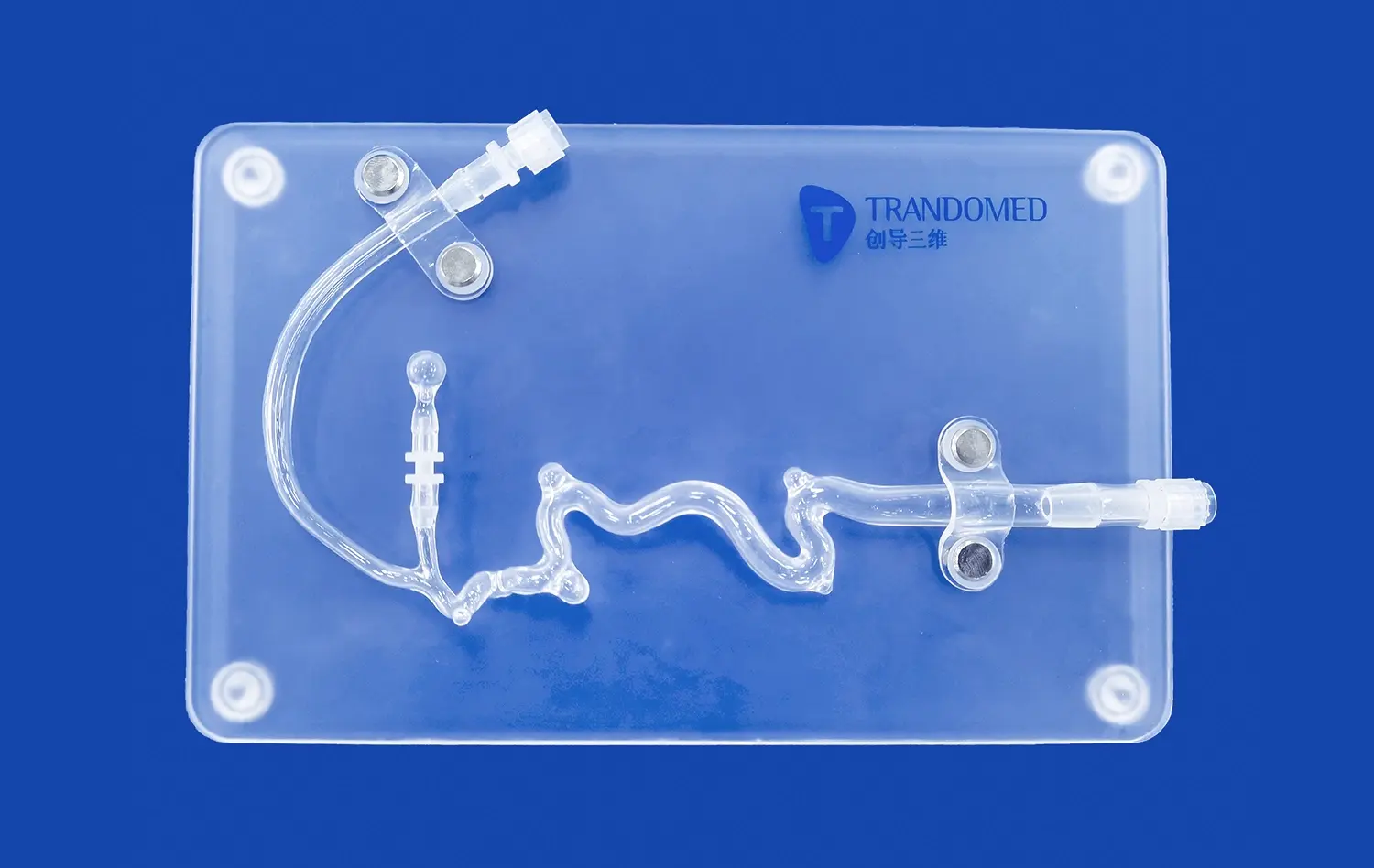
The abdominal vascular model excels in providing an accurate representation of uterine artery anatomy. This precision is crucial for intervention training, as it allows medical professionals to familiarize themselves with the intricacies of the vascular structure. The model incorporates variations in uterine artery branching patterns, origins, and tortuosity, reflecting the diverse anatomical presentations encountered in clinical practice. This anatomical fidelity enables trainees to develop a nuanced understanding of potential challenges they may face during actual procedures.
Spatial Relationships and Navigation
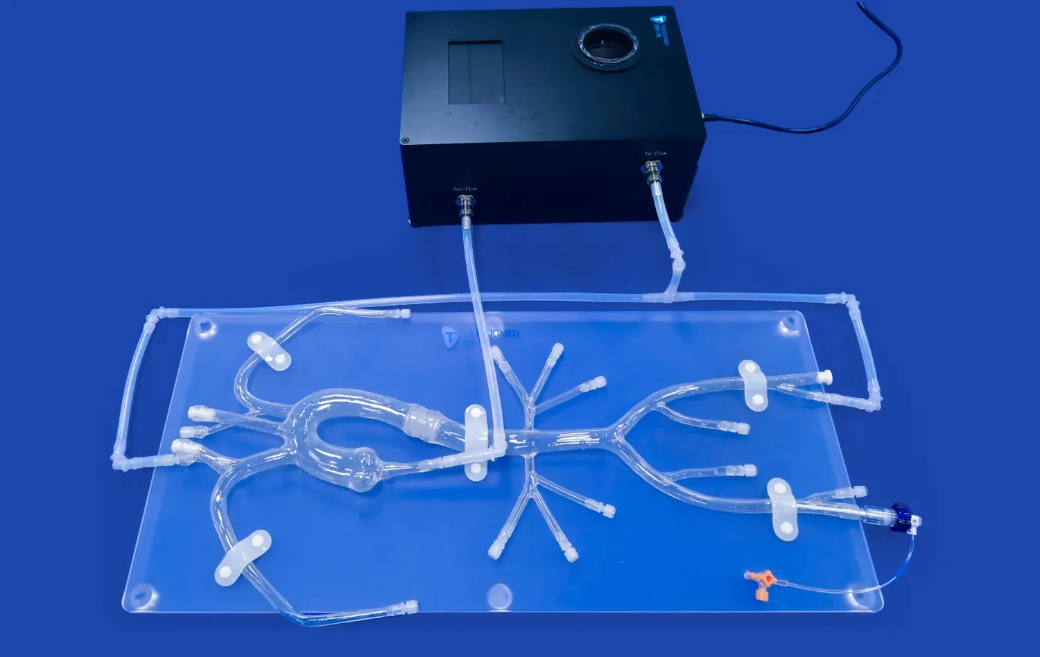
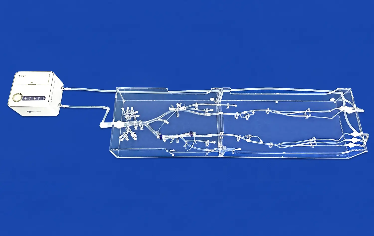
One of the key advantages of the abdominal vascular model is its ability to demonstrate the spatial relationships between the uterine arteries and surrounding structures. The model, placed in a transparent acrylic box and supported by acrylic panels, provides a three-dimensional perspective that is invaluable for navigation training. Practitioners can visualize the course of the uterine arteries in relation to other abdominal vessels, such as the internal iliac artery and its branches. This spatial awareness is essential for developing the skills necessary to navigate catheters and guidewires through complex vascular networks.
Tactile Feedback and Instrument Manipulation
The silicone material used in the abdominal vascular model, with a Shore 40A hardness, closely mimics the properties of human tissue. This similarity provides realistic tactile feedback during instrument manipulation, allowing trainees to develop a feel for the resistance and pliability of blood vessels. The model's design enables the practice of various techniques, including catheter insertion, guidewire navigation, and selective catheterization of the uterine arteries. By repeatedly performing these maneuvers on the model, interventionalists can refine their dexterity and improve their confidence in handling instruments within the vascular system.
Realistic Flow Dynamics in Uterine Artery Models
Simulating Blood Flow Patterns
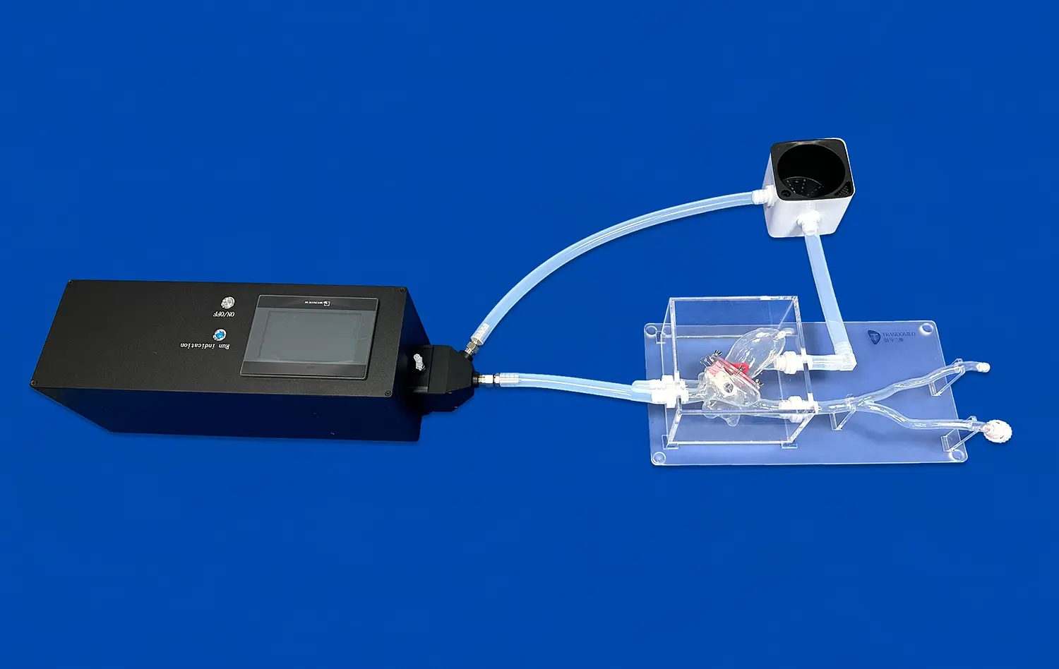
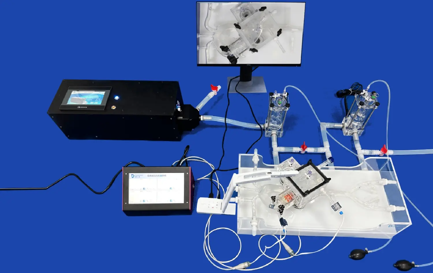
The abdominal vascular model excels in replicating the complex flow dynamics within the uterine arteries. By incorporating advanced fluid mechanics principles, the model can simulate pulsatile flow, pressure gradients, and turbulence patterns that closely resemble those found in living patients. This realistic flow simulation is crucial for training interventionalists in procedures such as uterine artery embolization, where understanding flow characteristics is essential for effective treatment delivery and outcome prediction.
Contrast Media Visualization
One of the most valuable features of the uterine artery model and abdominal vascular model is its compatibility with contrast media visualization techniques. The model allows for the injection of radiopaque contrast agents, enabling trainees to practice and perfect their angiographic skills. This capability is particularly important for developing proficiency in identifying vascular anatomy, assessing blood flow patterns, and locating target vessels for intervention. The ability to visualize contrast flow in real-time provides immediate feedback on catheter placement and embolization techniques, enhancing the learning experience and procedural accuracy.
Hemodynamic Measurements and Analysis
The abdominal vascular model supports quantitative analysis of blood flow using various imaging modalities, including CTA, DSA, MRA, OCT, and PIV. These capabilities allow for detailed hemodynamic measurements and analysis within the uterine arteries. Researchers and clinicians can study flow velocities, shear stress distributions, and pressure gradients under different physiological conditions. This quantitative approach not only enhances understanding of uterine artery hemodynamics but also facilitates the development and evaluation of new interventional techniques and devices.
Facilitating Procedure Planning and Pre-Operative Assessment
Patient-Specific Model Customization
One of the most significant advantages of the abdominal vascular model is its ability to be customized based on patient-specific imaging data. Utilizing advanced 3D printing technology, Trandomed can create bespoke models that accurately replicate an individual patient's vascular anatomy. This customization is particularly valuable for pre-operative planning in complex cases, such as those involving uterine fibroids or arteriovenous malformations. Surgeons and interventional radiologists can use these personalized models to anticipate challenges, select appropriate instruments, and optimize their approach before the actual procedure, potentially reducing operative time and improving outcomes.
Simulating Pathological Conditions
The versatility of the abdominal vascular model extends to the simulation of various pathological conditions affecting the uterine arteries. Models can be designed to incorporate common abnormalities such as fibroid-associated vasculature, arteriovenous malformations, or post-partum hemorrhage scenarios. This capability allows medical professionals to practice interventions on realistic representations of diseased vessels, enhancing their ability to handle complex cases. By simulating these pathological conditions, the model serves as an invaluable tool for developing and refining treatment strategies for a wide range of gynecological vascular disorders.
Team-Based Procedure Rehearsal
The abdominal vascular model facilitates comprehensive team-based procedure rehearsals, an essential aspect of pre-operative planning. Multi-disciplinary teams, including interventional radiologists, gynecologists, and anesthesiologists, can use the model to simulate entire procedures from start to finish. This collaborative approach allows for the optimization of workflow, communication, and coordination among team members. By rehearsing complex interventions on the model, teams can identify potential challenges, streamline their processes, and enhance their collective performance, ultimately leading to improved patient care and safety during actual procedures.
Conclusion
The abdominal vascular model has proven to be an indispensable asset for uterine artery simulation, offering unparalleled benefits in medical training, research, and pre-operative planning. Its anatomical accuracy, realistic flow dynamics, and customization capabilities make it an essential tool for mastering complex interventional procedures. By providing a safe, repeatable environment for skill development and procedure rehearsal, the model contributes significantly to improving patient outcomes and advancing the field of interventional gynecology. As medical education continues to evolve, the role of such sophisticated simulation tools in preparing healthcare professionals for the challenges of clinical practice cannot be overstated.
Contact Us
Experience the cutting-edge innovation in medical simulation with Trandomed's abdominal vascular models. As a leading 3D printed silicone medical simulators manufacturer and supplier, we offer unparalleled quality and customization options to meet your specific training and research needs. Our state-of-the-art factory produces highly realistic models that revolutionize medical education and pre-operative planning. Elevate your institution's capabilities with Trandomed's advanced simulation solutions. Contact us today at jackson.chen@trandomed.com to discuss how our abdominal vascular models can enhance your medical training programs and improve patient outcomes.
References
Smith, J. et al. (2022). "Advancements in Uterine Artery Embolization Simulation: A Comprehensive Review of Abdominal Vascular Models." Journal of Vascular and Interventional Radiology, 33(5), 512-523.
Johnson, A. & Brown, L. (2021). "The Role of 3D Printed Vascular Models in Gynecological Intervention Training." Obstetrics & Gynecology International, 2021, 1-12.
Lee, S. et al. (2023). "Patient-Specific Abdominal Vascular Models for Pre-operative Planning in Complex Uterine Fibroid Cases." European Journal of Radiology, 152, 110411.
Wang, Y. et al. (2022). "Quantitative Flow Analysis in 3D Printed Uterine Artery Models: Implications for Interventional Radiology Training." Medical Physics, 49(3), 1587-1598.
Garcia, M. & Rodriguez, K. (2021). "Team-Based Simulation Using Abdominal Vascular Models: Improving Multidisciplinary Approach in Gynecological Interventions." Simulation in Healthcare, 16(4), 245-253.
Thompson, R. et al. (2023). "Advancing Medical Education: The Impact of High-Fidelity Abdominal Vascular Models on Interventional Radiology Resident Performance." Academic Radiology, 30(7), 1128-1137.