Integrating Anatomical Accuracy into Curriculum
Enhancing 3D Visualization of Pulmonary Structures
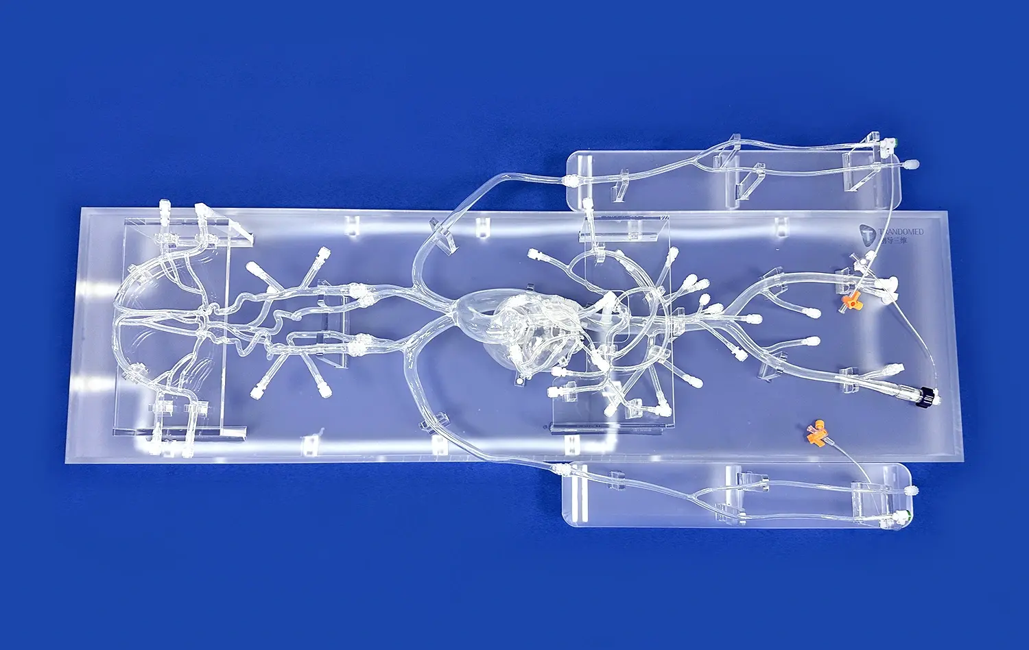
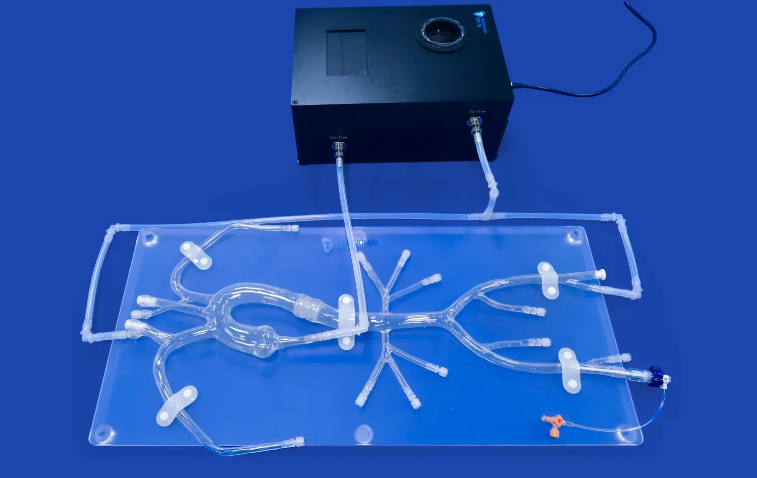
The pulmonary vein model serves as an invaluable asset in medical education by offering a tangible, three-dimensional representation of complex vascular structures. Unlike traditional 2D illustrations or digital renderings, these models allow students to physically interact with anatomically accurate representations. This hands-on approach significantly enhances spatial understanding and retention of pulmonary vein anatomy. The model's intricate details, including the left atrium, right atrium, and the four main branches of the pulmonary veins, provide a comprehensive view that's difficult to achieve through other educational methods.
Facilitating Comprehensive Understanding of Vascular Relationships
One of the key advantages of incorporating pulmonary vein models into medical curricula is their ability to demonstrate the intricate relationships between various vascular structures. The model's design, which includes the femoral vein, inferior vena cava, and both atria, allows students to visualize how these components interact within the cardiovascular system. This comprehensive approach helps learners grasp the bigger picture of cardiac anatomy, fostering a deeper understanding of how interventions in one area might affect other parts of the system.
Adapting to Various Learning Styles
The integration of pulmonary vein models caters to diverse learning preferences among medical students. Visual learners benefit from the model's clear, colorful representation of vascular structures. Kinesthetic learners thrive on the opportunity to physically manipulate the model, reinforcing their understanding through touch. Auditory learners can engage in discussions about the model's features, enhancing their comprehension through verbal explanations. This multi-sensory approach to learning ensures that the curriculum meets the needs of all students, regardless of their preferred learning style.
Demonstrating Interventional Techniques in Realistic Scenarios
Simulating Catheter Navigation and Placement
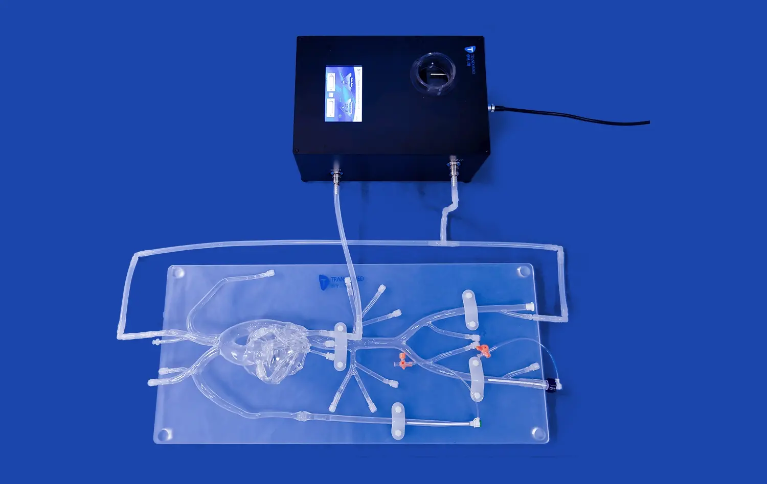
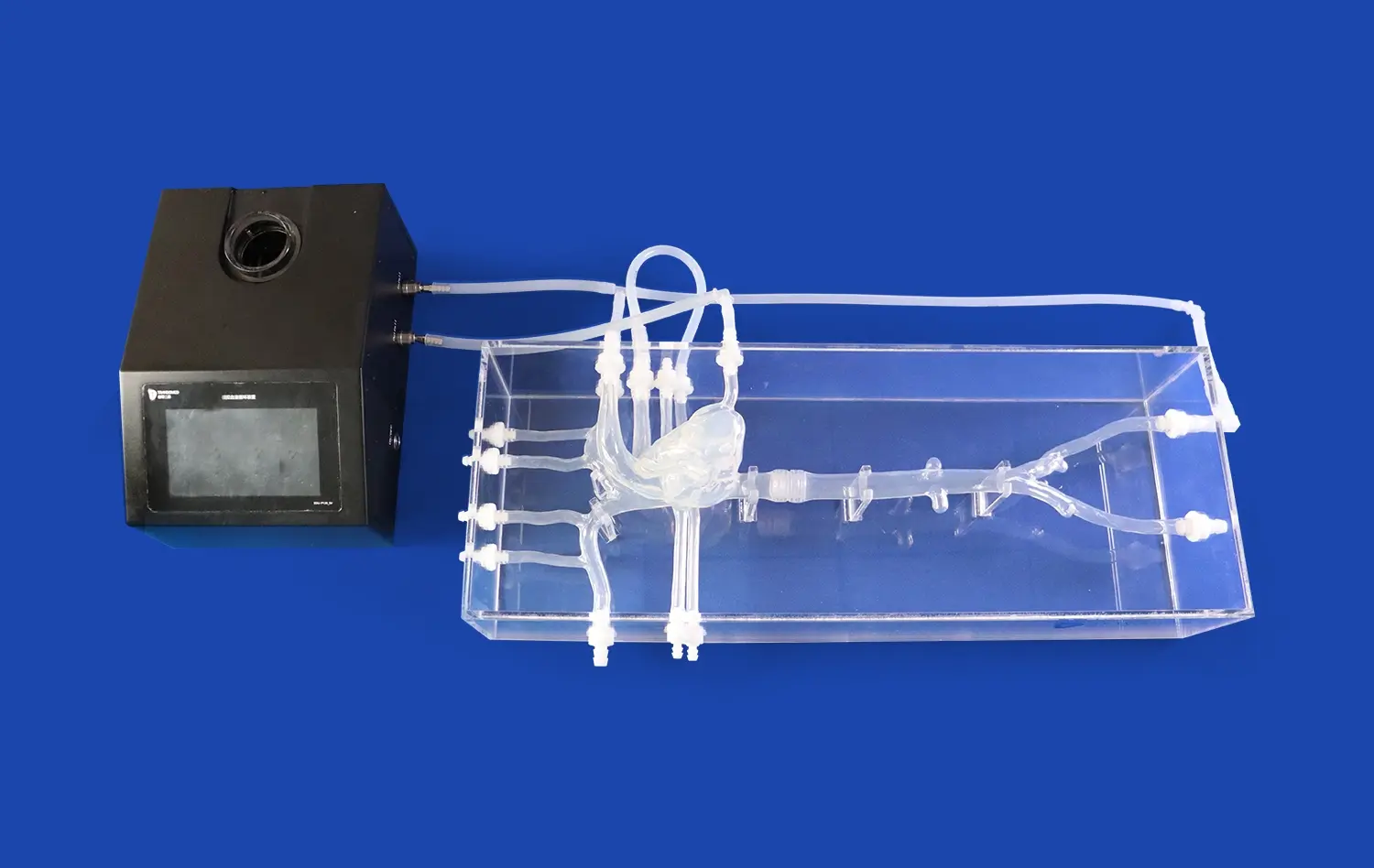
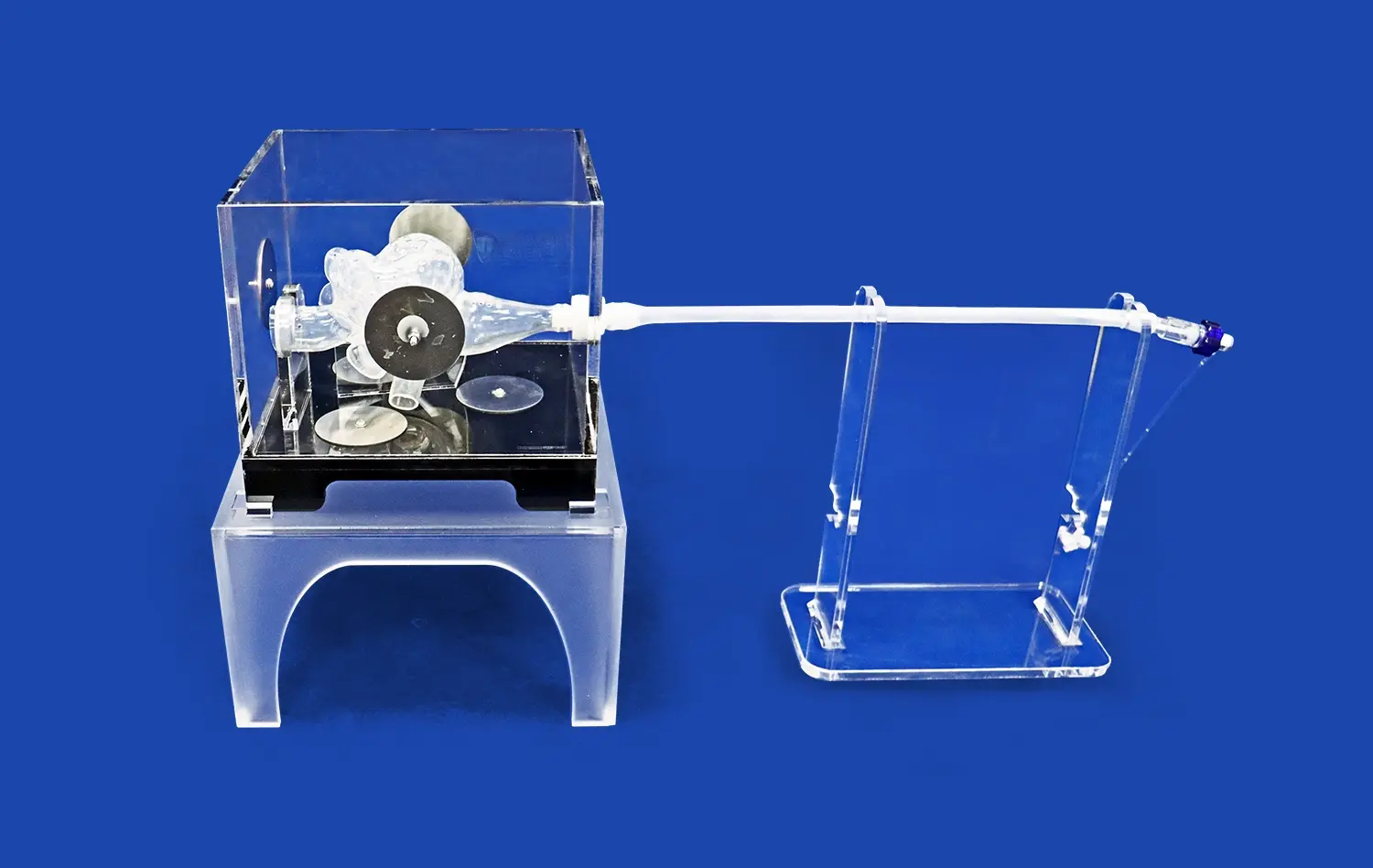
The pulmonary vein model excels in providing a realistic platform for practicing catheter navigation and placement. The model's design, featuring holes in the left and right atrium, allows for the insertion and manipulation of interventional instruments. This feature is particularly valuable for training in procedures such as pulmonary vein isolation, a common treatment for atrial fibrillation. Students and practitioners can refine their technique in guiding catheters through the vascular system, reaching target areas within the pulmonary veins with precision. The ability to practice these delicate maneuvers in a risk-free environment is invaluable for building confidence and competence before performing procedures on actual patients.
Practicing Advanced Imaging Techniques
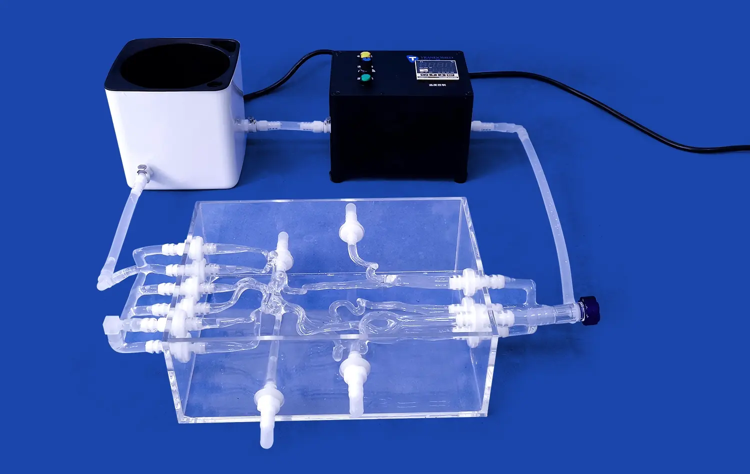
Another significant advantage of using pulmonary vein models in medical education is the opportunity to practice advanced imaging techniques. The model's compatibility with contrast agents allows for realistic simulation of angiography procedures. Students can learn to interpret real-time imaging results, understanding how different vascular structures appear under various imaging modalities. This hands-on experience with imaging techniques such as CTA, DSA, MRA, and OCT is crucial for developing the skills necessary for accurate diagnosis and intervention planning in clinical settings.
Exploring Pathological Variations
The versatility of pulmonary vein models extends to the representation of various pathological conditions. Educational institutions can utilize models that incorporate common abnormalities such as atrial septal defects, patent foramen ovale, or pulmonary vein stenosis. This feature allows students to familiarize themselves with a range of potential clinical scenarios they may encounter in practice. By interacting with these pathological variations, learners develop critical thinking skills and learn to adapt their approach based on individual patient anatomy and conditions.
Supporting Evidence-Based Training Programs
Enhancing Procedural Competency
Evidence-based training programs increasingly rely on pulmonary vein models to enhance procedural competency among medical professionals. These models provide a standardized platform for assessing and improving skills in cardiac interventions. Research has shown that repetitive practice on high-fidelity simulators leads to significant improvements in procedural performance and reduction in errors. By incorporating pulmonary vein models into their training regimens, medical institutions can objectively measure and track the progress of their trainees, ensuring they meet the required competency levels before advancing to patient care.
Facilitating Collaborative Learning
Pulmonary vein models serve as powerful tools for collaborative learning in medical education. They create opportunities for group discussions, peer-to-peer teaching, and interdisciplinary training sessions. For instance, a single model can be used to bring together students from various specialties - cardiology, radiology, and vascular surgery - fostering a comprehensive understanding of cardiac procedures from multiple perspectives. This collaborative approach not only enhances learning but also prepares students for the team-based nature of modern healthcare delivery.
Advancing Research and Innovation
Beyond their role in education, pulmonary vein models play a crucial part in advancing cardiac research and innovation. These models serve as testing platforms for new interventional devices and techniques, allowing researchers to evaluate the efficacy and safety of novel approaches before clinical trials. The ability to customize models based on specific patient data opens up possibilities for personalized medicine, enabling practitioners to plan and practice complex procedures tailored to individual patient anatomies. This integration of research and education ensures that medical institutions remain at the forefront of cardiovascular care, continually improving patient outcomes through evidence-based practices.
Conclusion
The adoption of pulmonary vein models in medical education represents a significant leap forward in preparing healthcare professionals for the complexities of cardiac care. These advanced simulation tools offer unparalleled opportunities for hands-on learning, skill development, and collaborative training. By integrating anatomical accuracy, demonstrating realistic interventional scenarios, and supporting evidence-based programs, pulmonary vein models have become indispensable in modern medical curricula. As the field of cardiovascular medicine continues to evolve, these innovative educational aids will play an increasingly vital role in shaping competent, confident, and skilled medical professionals ready to tackle the challenges of tomorrow's healthcare landscape.
Contact Us
Elevate your medical education program with Trandomed's state-of-the-art pulmonary vein models. As a leading 3D printed silicone medical simulators manufacturer and supplier, we offer customizable, high-fidelity solutions to meet your specific training needs. Experience the difference that anatomical accuracy and realistic simulation can make in your curriculum. Contact us today at jackson.chen@trandomed.com to explore how our advanced models can enhance your institution's cardiac education and research capabilities. Partner with Trandomed - your trusted factory for innovative medical simulation technology.