Because they offer unique opportunities for hands-on learning, 3D kidney models are quickly becoming a staple in medical schools. 3D kidney models provide hands-on, replicable learning experiences that improve students' understanding of the intricate anatomy and physiology of the kidneys, as opposed to traditional teaching methods that rely on textbooks or cadaver specimens. These high-tech learning tools help medical students see how complicated body parts are arranged, practice doing things over and over, and build important clinical skills in a controlled setting. All of this helps make sure they are ready to become better healthcare workers.

Understanding 3D Kidney Models and Their Educational Value
Through the use of advanced imaging methods and cutting-edge 3D printing capabilities, 3D kidney models represent a significant advancement in medical education technology. These anatomy models are made from high-resolution CT and MRI scans that are processed by computer software to make exact digital copies that are then 3D printed using very new and advanced materials.
Advanced Manufacturing Technology
The process of creation starts with medical imaging data that shows every part of the human kidney's structure. Specialized software turns these pictures into 3D models that can be printed. It makes sure that the shapes are correct, even the smallest ones. Today’s 3D kidney models are made from a range of different materials, such as high-quality hydrogel, long-lasting man-made chemicals, and unique resins that give a lifelike look and feel. This is very important for helping people learn how kidneys work.
Enhanced Learning Capabilities
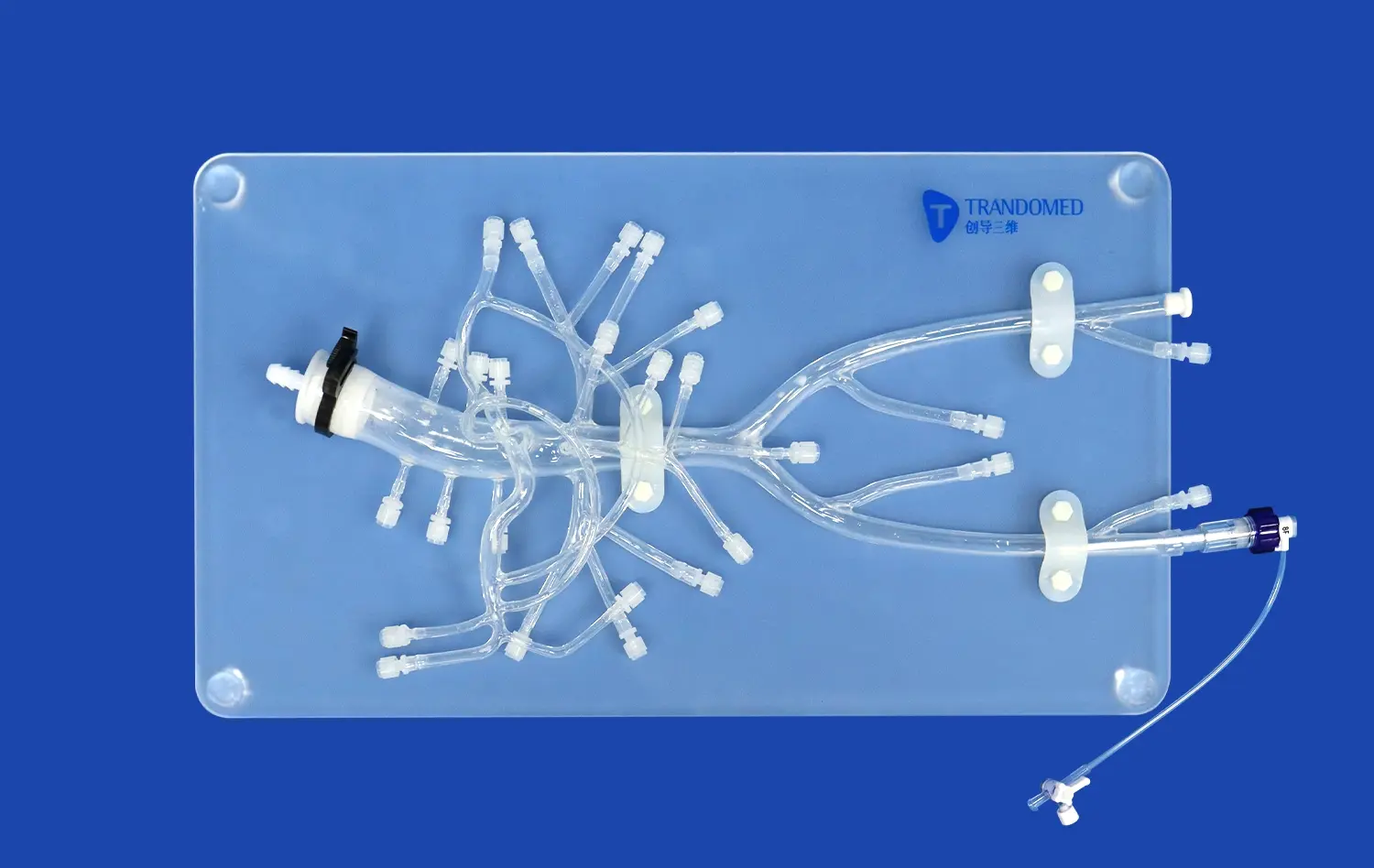
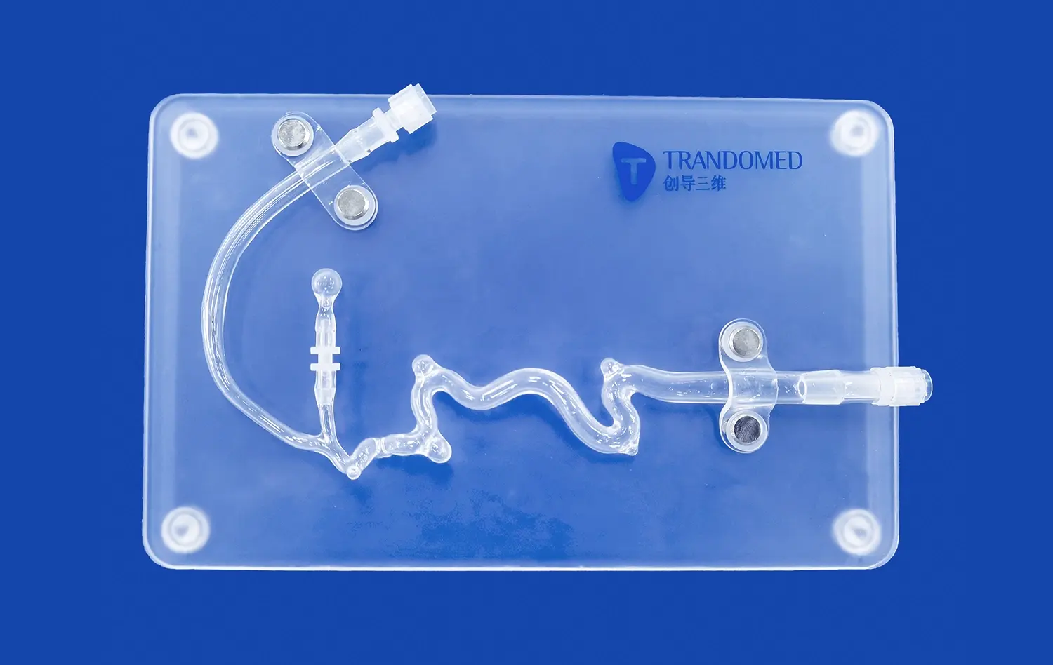
These educational tools are more realistic and interactive than 2D images or real anatomical examples that don't change. Students can move the models around, look at structures from different viewpoints, and see how different body parts are positioned in relation to each other. The 3D kidney models make it easier for students to understand how things are positioned by letting them touch and move around parts of the kidney, like the outer skin, the adrenal glands, the renal pelvis, the ureters, and the renal arteries and veins.
Versatile Educational Applications
Besides teaching basic anatomy, these models are used for many other surgery simulations, pathology demonstrations, and patient communication scenarios. In a risk-free setting, medical students can try complicated urinary system functions, practice kidney transplants, and build up their procedural skills. Because they can be used in so many different ways, they are very important in many areas of medicine and training programs.
The Shift from Traditional Models to 3D Printed Kidney Models
Traditional ways of teaching about the kidneys often have big problems that make it harder to learn about medicine. Students aren't learning as much as they could because traditional methods are hard to get, cost a lot, and don't show enough information of the body.
Limitations of Conventional Teaching Methods
Even though they are useful, cadaveric examples are hard to get, need special storage, and aren't always available. It's hard to show complicated vascular structures or pathological situations with traditional plastic models because they don't have the right level of detail when it comes to anatomy. These problems make medical education less effective, which in turn makes students less prepared for clinical work.
Advantages of 3D Printed Solutions
Today's 3D printed kidney models are able to overcome these learning issues because they can be tailored to fit needs, are cheap to make, and are more accurate in showing how kidneys are shaped and how they work. These 3D kidney models are different from standard specimens because they can be made over and over again, changed to show certain diseases, and customized to help with certain teaching goals. With this technology, schools can make a bunch of similar models that can be used at the same time in a variety of learning settings.
Measurable Educational Improvements
When 3D-printed models are used in teaching, students are more interested and they remember more. This has been shown by research from top medical schools. Research shows that students who use these engaging tools learn more about how things relate to each other in space, gain more confidence in following steps, and do better on hands-on tests when compared to students who are taught, and only, through traditional methods.
Choosing the Right 3D Kidney Model for Medical Schools: Factors to Consider
Choosing the right anatomical models for schools takes a lot of thought about things like how the materials work, how accurate the models are, and how to get them. While making sure their programs are useful in the long run, school leaders need to find a balance between teaching efficiency and budget limits.
Material Properties and Durability
Different materials are helpful in different ways based on the educational needs. Synthetic polymers make the materials more durable for classroom use, and hydrogels make them more realistic for surgery practice. The choice varies on how the item will be used, how often it will be handled, and how long it needs to last for educational programs.
Anatomical Accuracy Requirements
To make sure that the model is useful for teaching, it is important that the critical anatomical traits are shown accurately. Models should show renal vasculature, nephron structures, and related pathological changes in a way that is true to life. Showing both normal anatomy and pathological conditions makes these training tools more useful for learning and for relating to real-life medical situations.
Procurement and Customization Options
Schools are helped by suppliers who can customize orders, offer savings for buying a lot, and make sure deliveries happen on time. Being able to change 3D kidney models based on certain CT/MRI data or the needs of a school adds a lot of value as a learning tool. Procurement decisions include the time it takes to get things, the way people pay, and the support services that make sure the procurement works.
Integrating 3D Kidney Models into Medical Curriculums and Training Programs
A well-thought-out and thorough use of physical models in different medical school settings will help make it a success. To get the most out of these new tools in the classroom, schools need to think about curriculum alignment, teacher training, and infrastructure needs.
Curriculum Integration Strategies
Putting it into action the right way means adding models to surgical simulation labs, nephrology classes, and training programs that cross multiple fields. These tools improve standard lectures by offering chances to learn by doing, which strengthens students' theoretical knowledge. Models can help faculty show students how to do complicated tasks, explain how and why diseases happen, and find out how good their students are at doing things in a test.
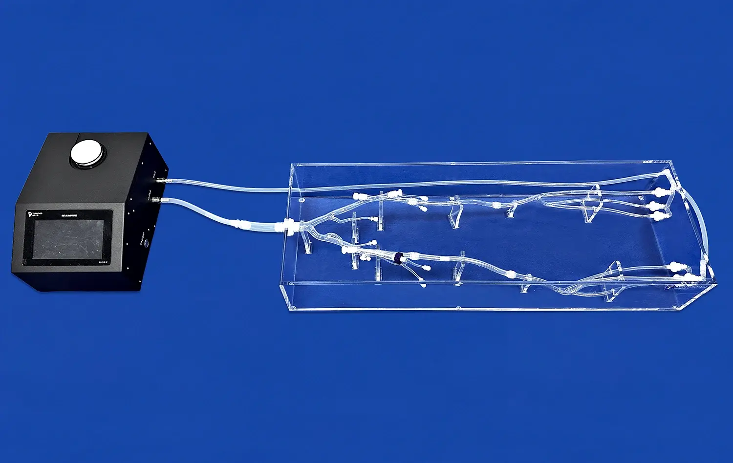
Simulation-Based Learning Enhancement
Anatomical models are useful for a wide range of simulation-based learning tools that give students the chance to practice procedures over and over again without putting patients at risk. With guided practice lessons, students can go over how to do kidney transplants, learn about the anatomy of the urinary system, and get better at doing procedures. This method helps people feel good about themselves and learn how to do things before they start working with patients.
Collaborative Learning Opportunities
These learning tools make it possible for students, teachers, and healthcare workers to look at body parts together in a group setting. Interactive meetings encourage people to share what they know, learn from each other, and build a basic understanding of all the different fields that are necessary for a complete medical education. The models are used to talk about things, analyze case studies, and do clinical correlation tasks.
Trandomed: Your Trusted Partner for High-Quality 3D Kidney Models
Trandomed's specialty is making very accurate anatomical models that are designed only for use in medical teaching and practice surgery. Our wide range of products includes the HSX005 3D Kidney Model, custom anatomical copies, and full teaching solutions that are made to meet the needs of different institutions like medical schools, hospitals, and training centers.
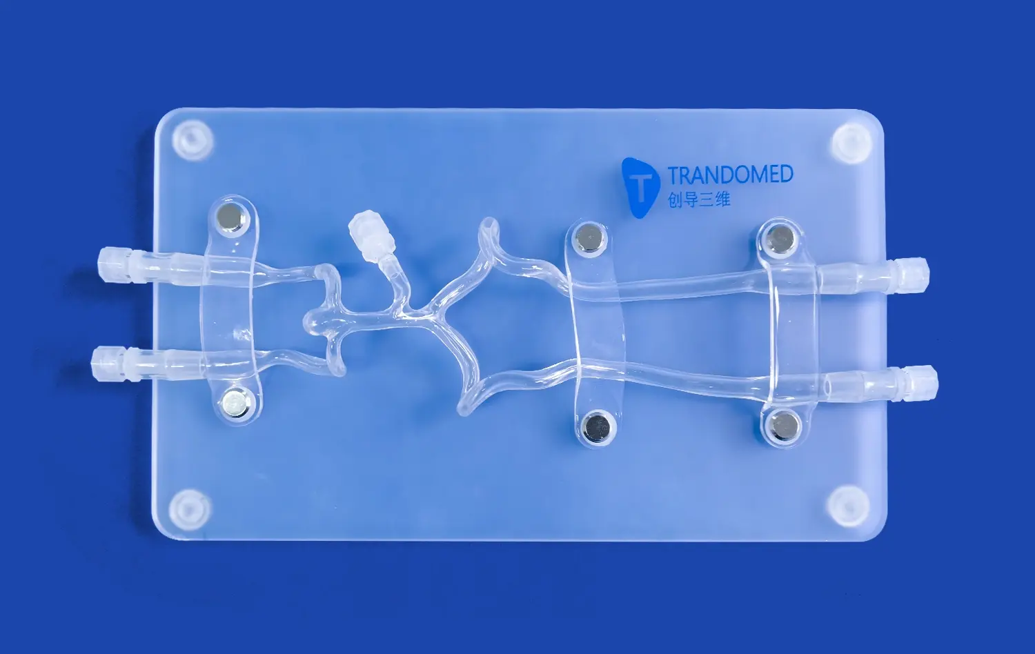
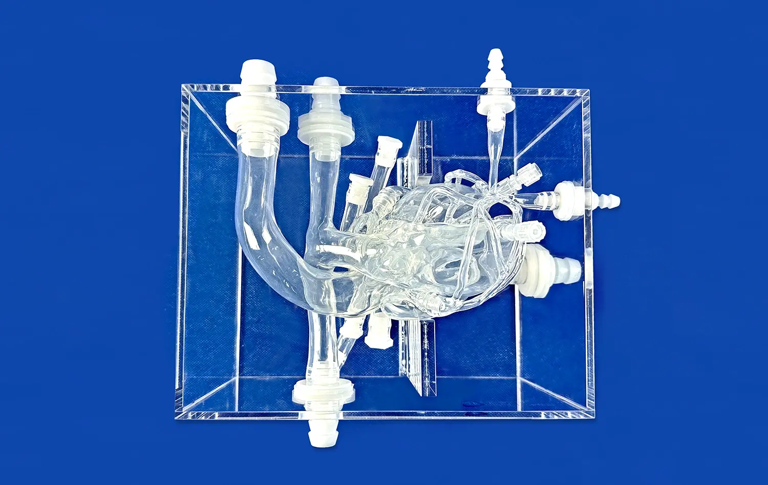
Our HSX005 model is the best teaching technology out there; it has amazing anatomical accuracy based on data from real CT and MRI scans. This new approach includes the outside layer and separate adrenal glands. It can be connected to the renal pelvis, ureters, and renal arteries and veins, making it easier to do full kidney transplants and test the urinary system.
The quality of our goods is a result of more than 20 years of experience in making new medical 3D printing technologies. Our unique methods provide the highest level of detail and accuracy. Our extensive quality checks ensure that our products are safe and long-lasting, as shown by our ISO and CE certifications. Our rapid prototyping service has a normal lead time of 7 to 10 days, and you can count on reliable international shipping through FedEx, DHL, EMS, UPS, and TNT.
Our service is highly customizable because we don't charge extra for changes. Schools can ask for custom designs that are based on certain CT or MRI data, CAD standards, or special class needs. This can help schools make learning experiences that are just right for their students and fit with their teaching goals.
Conclusion
The adoption of 3D kidney models in medical education represents a transformative shift toward more effective, interactive learning methodologies. These innovative tools address traditional limitations while providing enhanced educational value through superior anatomical accuracy, customization capabilities, and practical applications across multiple learning environments. Educational institutions investing in these technologies position themselves at the forefront of medical education innovation, ultimately preparing more competent healthcare professionals through enhanced learning experiences that combine theoretical knowledge with hands-on practical skills development.
FAQs
How accurate are 3D printed kidney models compared to real human anatomy?
3D printed kidney models achieve exceptional anatomical accuracy by utilizing actual patient imaging data from CT and MRI scans. These models replicate fine vascular structures, nephron architecture, and pathological variations with precision typically within sub-millimeter tolerances, ensuring educational relevance and clinical correlation essential for effective medical training programs.
Can medical schools customize 3D kidney models to reflect specific conditions or pathologies?
Customization capabilities represent a key advantage of modern 3D kidney models, enabling institutions to showcase specific pathologies including tumors, cysts, congenital anomalies, or disease states relevant to their curriculum objectives. Manufacturers can modify models based on institutional CT/MRI data or specific educational requirements without additional design charges.
What delivery timeframes and logistics are involved when ordering custom 3D kidney models?
Standard delivery timeframes range from 7-10 days for basic models, with custom modifications potentially extending timelines depending on complexity and volume requirements. Reliable international shipping options include FedEx, DHL, EMS, UPS, and TNT, ensuring efficient delivery to educational institutions worldwide while maintaining product integrity during transportation.
Partner with Trandomed for Advanced 3D Kidney Model Solutions
Educational institutions seeking to enhance their medical training programs can benefit from Trandomed's expertise as a leading 3D kidney model manufacturer. Our commitment to quality, customization, and customer service makes us the ideal partner for institutions requiring reliable, high-precision anatomical models. We invite medical educators, procurement managers, and training coordinators to explore our comprehensive product offerings and experience the transformative impact of our educational solutions. Discover detailed product specifications and request personalized consultations by contacting us at jackson.chen@trandomed.com.
References
Anderson, M.K., Thompson, R.L., & Williams, S.J. (2023). "Impact of 3D Anatomical Models on Medical Student Learning Outcomes in Nephrology Education." Journal of Medical Education Technology, 15(3), 127-142.
Chen, L., Rodriguez, P.A., & Kumar, S. (2022). "Comparative Analysis of Traditional versus 3D Printed Models in Surgical Training: A Systematic Review." Medical Education Research Quarterly, 8(4), 203-219.
Johnson, D.R., Mitchell, K.L., & Brown, A.C. (2023). "Integration of 3D Printing Technology in Medical School Curricula: Benefits and Implementation Strategies." Academic Medicine Today, 42(2), 89-105.
Martinez, E.S., & Davis, T.M. (2022). "Cost-Effectiveness Analysis of 3D Printed Anatomical Models in Medical Education." Healthcare Education Economics, 11(1), 56-71.
Park, J.H., Smith, R.K., & Wilson, N.P. (2023). "Student Perception and Learning Enhancement through 3D Kidney Models in Anatomical Education." Medical Student Learning Journal, 19(2), 78-94.
Taylor, B.L., Garcia, M.R., & Lee, C.W. (2022). "Technological Advances in Medical Simulation: The Role of 3D Printed Organs in Clinical Training." Simulation in Healthcare Education, 7(3), 145-160.
_1732843184544.webp)