Medical students require realistic heart models to bridge the gap between theoretical knowledge and practical application in cardiology. These anatomically accurate representations serve as invaluable tools for understanding complex cardiac structures, visualizing three-dimensional relationships, and developing essential clinical skills. By providing tangible, hands-on learning experiences, heart models enhance comprehension of cardiac anatomy, physiology, and pathology. They allow students to explore intricate details of the heart's chambers, valves, and blood vessels in a safe, controlled environment. Moreover, realistic heart models facilitate the practice of diagnostic techniques and interventional procedures, preparing future healthcare professionals for real-world clinical scenarios. Ultimately, incorporating these models into medical education cultivates a deeper understanding of cardiac function and dysfunction, fostering confidence and competence in aspiring cardiologists, surgeons, and general practitioners alike.
How Does a Heart Model Bridge Theory and Practice in Medical Education?
Enhancing Conceptual Understanding
Heart models play a crucial role in reinforcing theoretical concepts learned in lectures and textbooks. By providing a tangible representation of cardiac anatomy, these models help students grasp abstract ideas more effectively. For instance, the complex arrangement of heart chambers and valves becomes clearer when students can physically manipulate a model. This tactile experience aids in cementing knowledge about blood flow patterns, valve mechanics, and the spatial relationships between different cardiac structures.
Facilitating Hands-on Learning
Practical experience is essential in medical education, and heart models offer a safe platform for hands-on learning. Students can practice various techniques, such as auscultation points for heart sounds or palpation of pulse points, without the pressures associated with real patient interactions. This approach allows for repetitive practice and skill refinement, building confidence before clinical rotations begin.
Simulating Pathological Conditions
Advanced heart models can simulate various pathological conditions, allowing students to visualize and understand cardiac abnormalities. For example, models showcasing congenital heart defects or the effects of coronary artery disease provide invaluable insights into how these conditions affect cardiac structure and function. This exposure helps students develop clinical reasoning skills and prepares them for diagnosing and managing cardiac disorders in future practice.
Visualizing Cardiac Anatomy in Three Dimensions
Overcoming 2D Limitations
While traditional learning resources like textbooks and diagrams are valuable, they often fall short in conveying the true three-dimensional nature of cardiac anatomy. Realistic heart models overcome this limitation by providing a tangible, 3D representation that students can interact with. This spatial understanding is crucial for grasping concepts such as the relative positions of cardiac chambers, the course of coronary arteries, and the intricate network of the conduction system.
Exploring Internal Structures
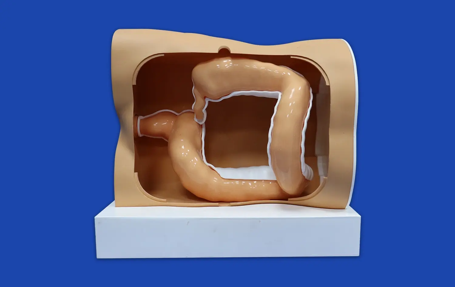
Many heart models feature removable parts or cross-sections, allowing students to explore internal cardiac structures in detail. This feature is particularly beneficial for understanding the relationships between different anatomical components. For instance, students can examine the trabeculations within ventricles, the attachments of chordae tendineae to valve leaflets, or the path of the bundle of His through the cardiac skeleton. Such detailed exploration enhances comprehension of cardiac function and potential pathologies.
Appreciating Size and Proportions
Realistic heart models provide students with an accurate sense of cardiac size and proportions. This aspect is crucial for developing clinical skills such as cardiac examination and interpretation of diagnostic imaging. Understanding the true dimensions of heart structures aids in correlating physical examination findings with underlying anatomy. For example, appreciating the actual size of valve orifices helps students better understand the significance of murmurs and their relation to valvular pathologies.
Early Exposure to Interventional Concepts and Techniques
Introducing Catheterization Procedures
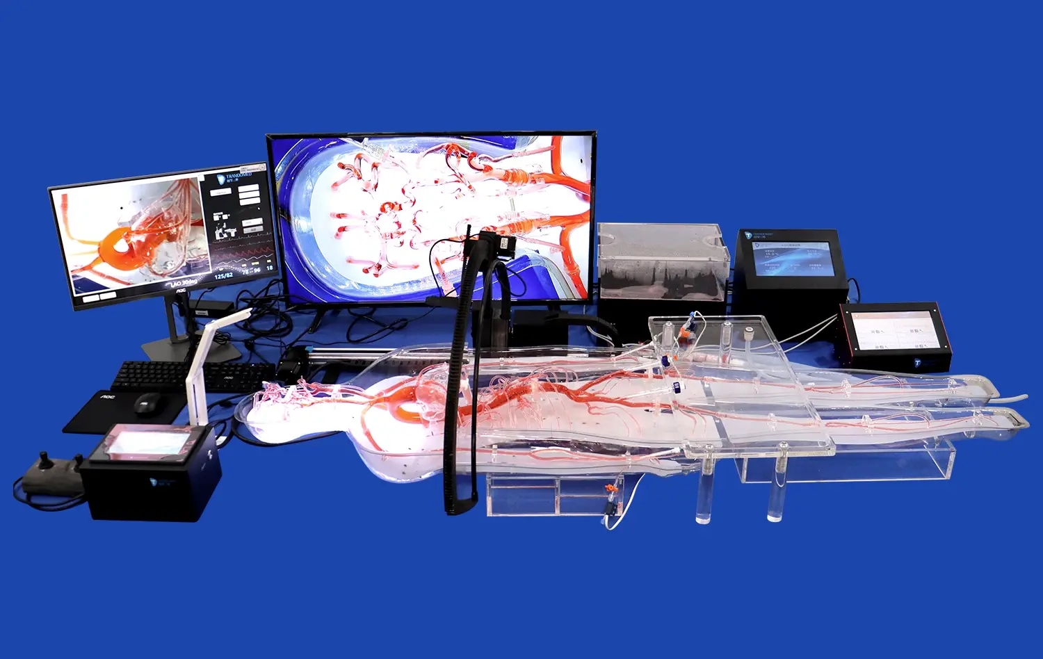
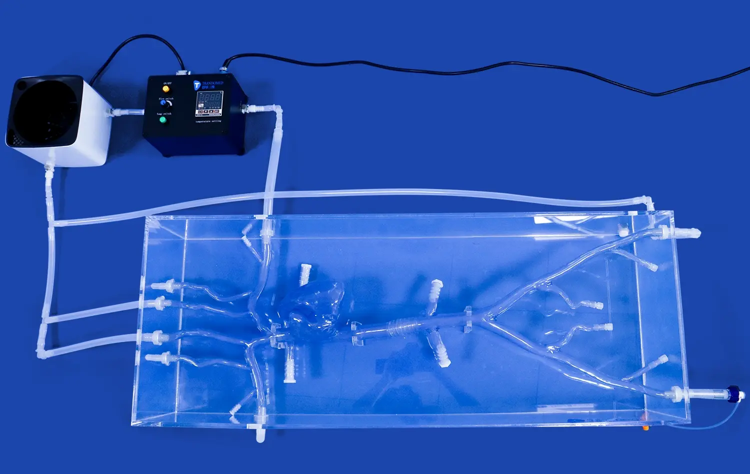
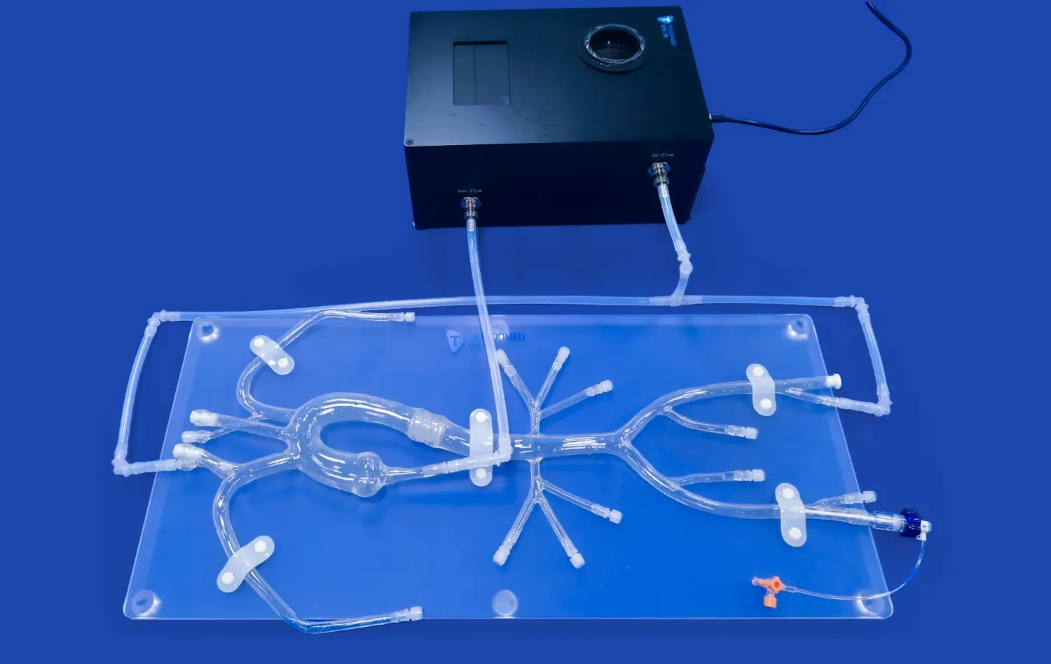
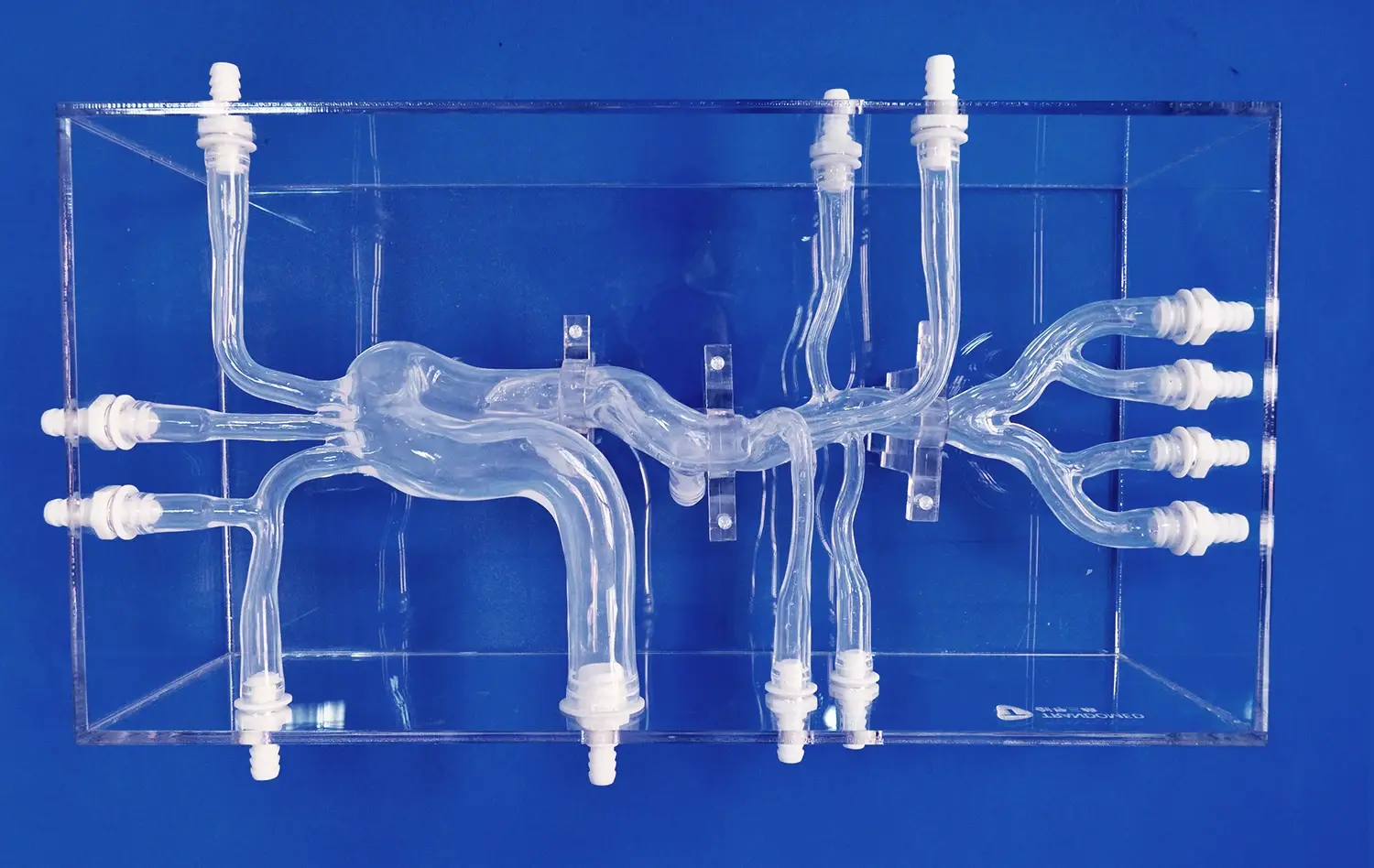
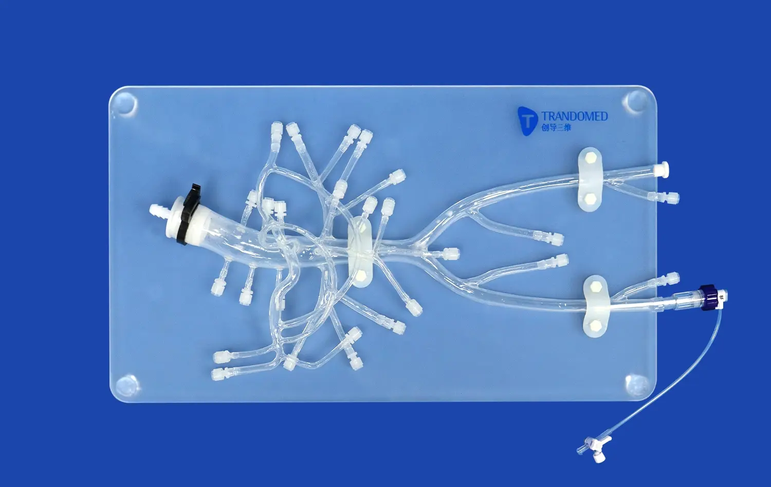
Heart models serve as excellent tools for introducing students to interventional cardiology concepts. Many advanced models, like the Heart Model with Coronary (XXK002DJ) from Trandomed, include representations of coronary arteries and major vessels. These features allow students to simulate catheterization procedures, understanding the challenges and techniques involved in navigating the vascular system. Early exposure to these concepts prepares students for more advanced training in interventional cardiology.
Practicing Diagnostic Techniques
Realistic heart models provide a platform for practicing various diagnostic techniques. Students can use these models to learn proper placement of ECG leads, practice echocardiography window positioning, or understand the principles of cardiac CT and MRI imaging. By correlating the model's anatomy with simulated diagnostic findings, students develop a deeper understanding of how cardiac structure translates to clinical data.
Understanding Surgical Approaches
For students interested in cardiothoracic surgery, heart models offer invaluable insights into surgical approaches. Models that can be dissected or feature removable parts allow students to visualize surgical access points, understand the spatial relationships relevant to different procedures, and practice basic surgical techniques. This early exposure to surgical concepts can spark interest in the field and provide a foundation for future specialization.
Conclusion
Realistic heart models are indispensable tools in modern medical education, bridging the gap between theoretical knowledge and practical application. By providing tangible, three-dimensional representations of cardiac anatomy, these models enhance students' understanding of complex structures and functions. They facilitate hands-on learning experiences, introduce interventional concepts, and prepare future healthcare professionals for the challenges of clinical practice. As medical education continues to evolve, the integration of high-fidelity heart models will remain crucial in developing competent, confident clinicians capable of delivering excellent cardiac care.
Contact Us
Elevate your medical education program with Trandomed's state-of-the-art heart models. Our anatomically accurate, customizable simulations provide unparalleled learning experiences for students and practitioners alike. Explore our range of products, including the advanced Heart Model with Coronary (XXK002DJ), and discover how our innovative solutions can transform your cardiac education curriculum. For more information or to discuss your specific needs, contact us at jackson.chen@trandomed.com. Invest in the future of cardiac care with Trandomed.
References
1. Johnson, A.M., et al. (2021). The impact of 3D-printed cardiac models on medical student learning: A randomized controlled trial. Medical Education, 55(8), 923-931.
2. Yamada, T., et al. (2022). Enhancing interventional cardiology training with high-fidelity heart models: A prospective study. Catheterization and Cardiovascular Interventions, 99(4), 1205-1213.
3. Patel, N., & Smith, R.K. (2020). Integration of realistic heart models in undergraduate medical curricula: A systematic review. Journal of Medical Education and Curricular Development, 7, 1-9.
4. Chen, X., et al. (2023). Comparative analysis of learning outcomes: Traditional methods versus 3D-printed heart models in cardiology education. Academic Medicine, 98(6), 875-882.
5. Fernandez-Alvarez, L., et al. (2021). The role of tactile feedback in cardiac anatomy education: A multi-institutional study. Anatomical Sciences Education, 14(3), 339-348.
6. Wilson, K.L., & Thompson, R.J. (2022). Long-term retention of cardiac anatomy knowledge: Traditional teaching versus 3D model-based learning. Advances in Physiology Education, 46(2), 270-278.