How Vascular Models Support High-Resolution OCT and MRA Imaging
Anatomical Precision for Enhanced Visualization
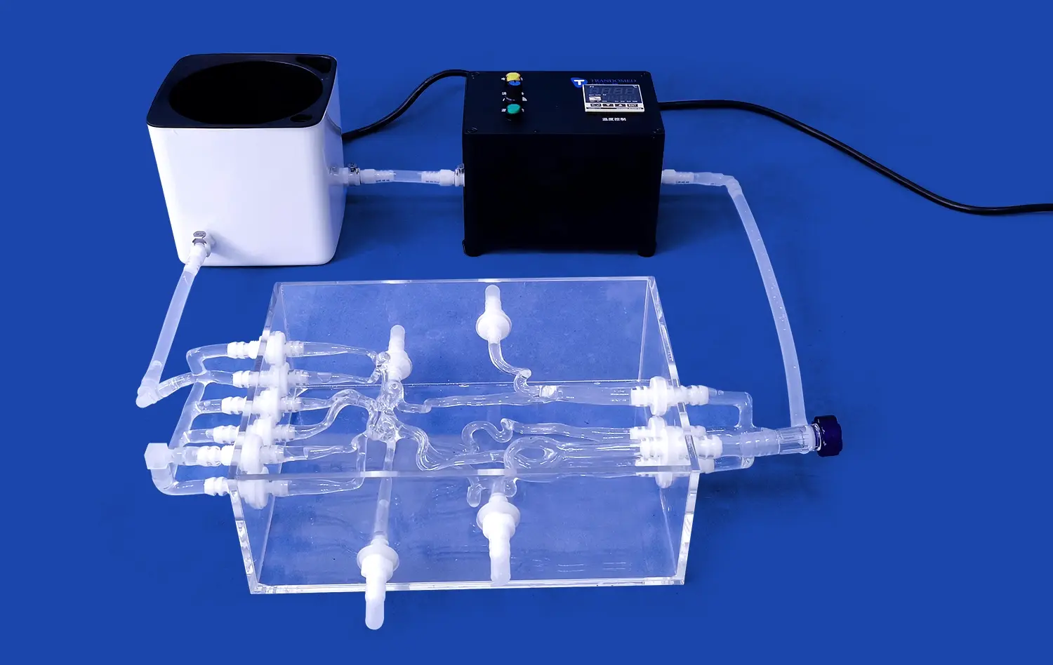
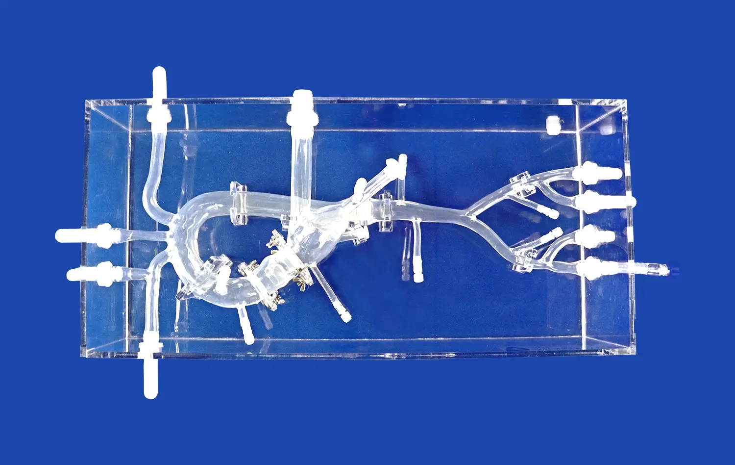
Abdominal vascular models play a pivotal role in supporting high-resolution OCT and MRA imaging by offering anatomically precise replicas of complex vascular structures. These models, crafted using advanced 3D printing technology, capture intricate details of blood vessels, including the celiac trunk, hepatic artery, and renal arteries. The level of detail in these simulators allows researchers to visualize minute vascular features that might be challenging to observe in live subjects due to motion artifacts or anatomical variations.
The anatomical accuracy of these models extends to replicating vessel wall thickness, lumen diameters, and branching patterns. This precision is crucial for OCT imaging, which requires high spatial resolution to detect subtle changes in vessel wall morphology. Similarly, for MRA, the models provide clear, static representations of vascular anatomy, enabling researchers to fine-tune imaging parameters without the complexities introduced by physiological motion or blood flow dynamics.
Optimizing Imaging Parameters
Researchers utilize abdominal vascular models to optimize imaging parameters for both OCT and MRA modalities. These models serve as consistent benchmarks for adjusting settings such as contrast, resolution, and scan protocols. For OCT, the models allow for experimentation with different light sources, probe designs, and image processing algorithms to enhance the clarity and depth of vascular imaging. In MRA studies, the models facilitate the optimization of pulse sequences, contrast agent protocols, and image reconstruction techniques.
By using standardized vascular phantoms, researchers can systematically evaluate the impact of various imaging parameters on image quality and diagnostic accuracy. This process is essential for developing robust imaging protocols that can be reliably translated to clinical applications, ensuring that the full potential of OCT and MRA technologies is realized in vascular diagnostics and interventional planning.
Simulating Pathological Conditions
Advanced abdominal vascular models go beyond normal anatomy, incorporating simulations of various pathological conditions. These can include stenoses, aneurysms, and irregular vessel wall thickening. By integrating these pathological features into the models, researchers can assess the sensitivity and specificity of OCT and MRA in detecting and characterizing vascular abnormalities.
This capability is particularly valuable for validating new imaging techniques or contrast agents designed to highlight specific vascular pathologies. Researchers can create controlled scenarios to test the limits of imaging technologies in differentiating between normal and diseased vessels, evaluating the accuracy of quantitative measurements, and developing automated image analysis algorithms for disease detection and classification.
Standardizing Imaging Protocols Through Reproducible Models
Consistent Baseline for Comparative Studies
Abdominal vascular models provide a consistent baseline for comparative studies across different imaging modalities and research centers. By using identical or highly similar models, researchers can establish standardized protocols for image acquisition and analysis. This standardization is crucial for ensuring that results from OCT and MRA studies are comparable and reproducible, regardless of the specific equipment or institution involved.
The use of these models allows for the creation of reference datasets that can be shared among researchers, facilitating collaborative efforts and meta-analyses. This approach is particularly beneficial in multi-center trials or when validating new imaging technologies against established standards. The consistency offered by these models helps in isolating variables related to imaging technology or technique, rather than anatomical variations between subjects.
Protocol Development and Validation
Researchers leverage abdominal vascular models to develop and validate imaging protocols for both OCT and MRA. These models serve as ideal test subjects for iterative refinement of scanning parameters, contrast agent administration techniques, and image reconstruction algorithms. By using models with known dimensions and features, researchers can quantitatively assess the accuracy and precision of their imaging protocols.
This process is essential for establishing best practices in vascular imaging. For instance, in OCT studies, researchers can determine optimal pullback speeds, frame rates, and focusing techniques to maximize image quality and diagnostic value. In MRA, the models help in optimizing sequence parameters, timing of contrast agent injection, and post-processing techniques to enhance vessel visualization and reduce artifacts.
Quality Assurance and Calibration
Abdominal vascular models play a crucial role in quality assurance and calibration of imaging systems. Regular use of these standardized models allows researchers and clinicians to verify the performance of OCT and MRA equipment over time, ensuring consistent image quality and accuracy. This is particularly important in longitudinal studies or when comparing results across different time points or imaging centers.
The models serve as phantoms for routine calibration checks, helping to identify and correct any drift in system performance. By periodically imaging these known structures, researchers can detect subtle changes in imaging parameters that might affect study outcomes. This proactive approach to quality control enhances the reliability of research findings and supports the translation of imaging techniques from bench to bedside.
Improving Experimental Reliability in Preclinical Studies
Reducing Variability in Imaging Studies
Abdominal vascular models significantly contribute to reducing variability in preclinical imaging studies. Unlike live animal models or human subjects, these synthetic models eliminate biological variability, allowing researchers to focus solely on the performance of imaging technologies and techniques. This reduction in confounding factors is crucial for isolating the effects of specific imaging parameters or interventions.
By using standardized models, researchers can conduct repeated measurements under identical conditions, enhancing the statistical power of their studies. This is particularly valuable in the early stages of technology development or when comparing the performance of different imaging modalities. The consistency provided by these models helps in establishing baseline performance metrics and identifying subtle improvements in imaging capabilities.
Facilitating Longitudinal Studies
Abdominal vascular models are invaluable tools for conducting longitudinal studies in preclinical research. Unlike biological specimens, these models do not change over time, allowing for consistent imaging over extended periods. This stability is crucial for evaluating the long-term performance of imaging technologies, assessing the durability of contrast agents, or studying the effects of repeated imaging on image quality.
Researchers can use the same model to track improvements in imaging techniques or to compare the performance of different imaging systems over time. This approach is particularly useful in developing and refining algorithms for image analysis, as it provides a consistent dataset for training and validation. The ability to conduct longitudinal studies with minimal variability accelerates the development and validation of new imaging technologies and protocols.
Ethical Considerations and Resource Efficiency
The use of abdominal vascular models in preclinical studies addresses important ethical considerations by reducing the need for animal experiments in the early stages of research. These models allow for extensive testing and optimization of imaging protocols without the ethical complexities associated with animal or human subject research. This approach aligns with the principles of the 3Rs (Replacement, Reduction, and Refinement) in scientific research.
Furthermore, vascular models offer significant resource efficiency. They can be used repeatedly without degradation, reducing the costs and logistical challenges associated with biological specimens. This efficiency allows researchers to conduct more extensive and thorough validation studies, potentially accelerating the translation of new imaging technologies to clinical applications. The reproducibility and consistency of these models also mean that fewer experimental iterations may be needed to achieve statistically significant results, further conserving research resources.
Conclusion
Abdominal vascular models have become indispensable tools for researchers validating OCT and MRA technologies. These advanced simulators offer unparalleled consistency, reproducibility, and anatomical accuracy, enabling the standardization of imaging protocols and enhancing the reliability of preclinical studies. By providing a controlled environment for high-resolution imaging, these models facilitate the optimization of imaging parameters, reduce experimental variability, and support longitudinal research efforts. As the field of vascular imaging continues to evolve, the role of these sophisticated models in advancing diagnostic and interventional techniques remains crucial, paving the way for improved patient care and more effective vascular health management strategies.
Contact Us
At Trandomed, we're at the forefront of 3D-printed medical simulation technology. As a leading high-fidelity abdominal vascular models manufacturer and supplier, we offer customizable solutions to meet your specific research needs. Our state-of-the-art factory produces anatomically accurate models that support cutting-edge OCT and MRA validation studies. Experience the difference that precision-engineered vascular simulators can make in your research. Contact us today at jackson.chen@trandomed.com to discuss how our advanced abdominal vascular model products can elevate your imaging research and medical training programs.
_1734504221178.webp)
_1734507815464.webp)
_1732866687283.webp)