Why the Circle of Willis Brain Model Matters in Neurosurgical Training?
2026-01-29 09:00:03
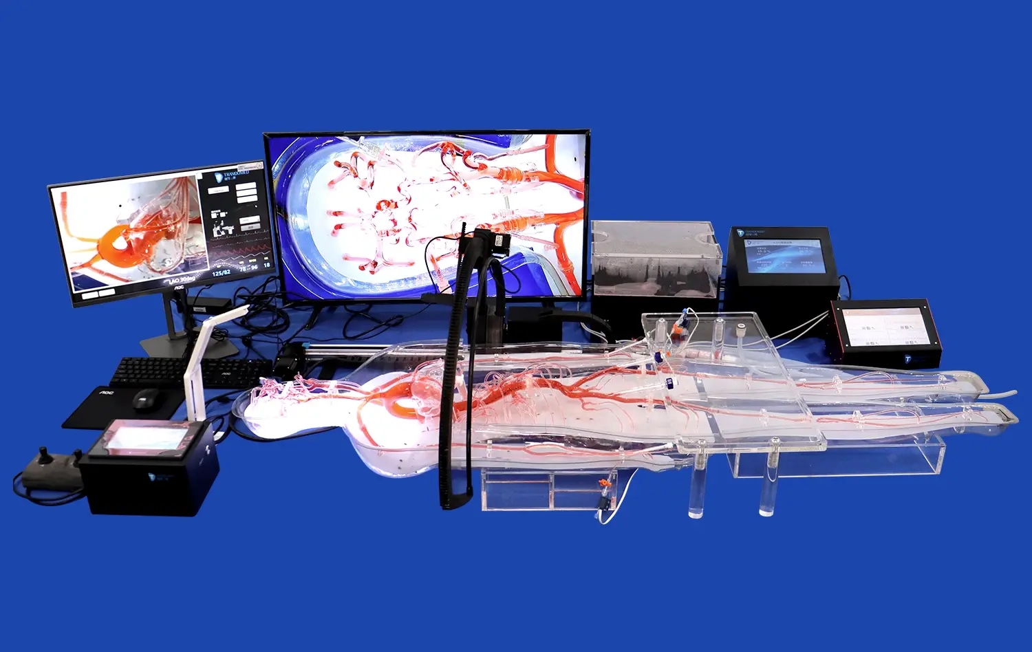
Because it accurately depicts the complex network of blood vessels that carry blood to the brain, the circle of Willis brain model is an essential teaching tool in neurosurgery. This part of the body is a very important safety device because it provides collateral circulation that keeps the brain from getting ischemic damage when blood vessels get sick or damaged. Only 20 to 25 percent of people have a full Circle of Willis, so it's important to know all of its differences in order to plan surgery. Neurosurgeons, doctors, and medical students can practice complex invasive procedures in a controlled setting thanks to advanced 3D-printed models. This lowers the risk to patients and speeds up skill development. These modeling tools bridge the gap between academic knowledge and clinical application by letting you touch real diseases like aneurysms and stenosis lesions. This improves the safety and results of surgery.
Understanding the Circle of Willis Brain Model: Anatomy and Function
In the human body, the cerebral artery circle is one of the most complex circulatory systems. Accurate modeling through computer models has changed the way neurosurgery is taught. The brain gets a steady flow of blood through this network, which is made up of many vessels that are all linked to each other.
Anatomical Components and Their Clinical Significance
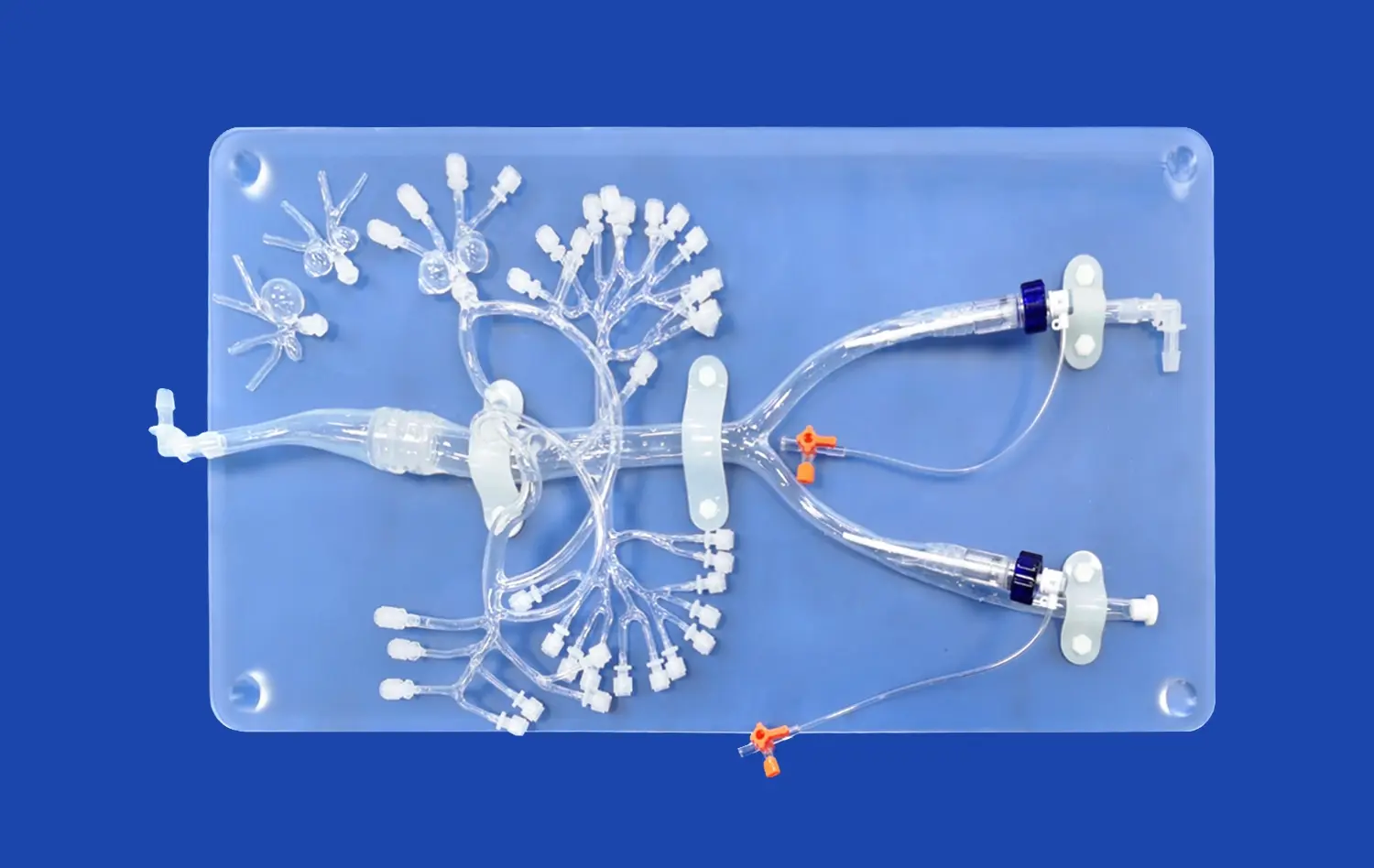
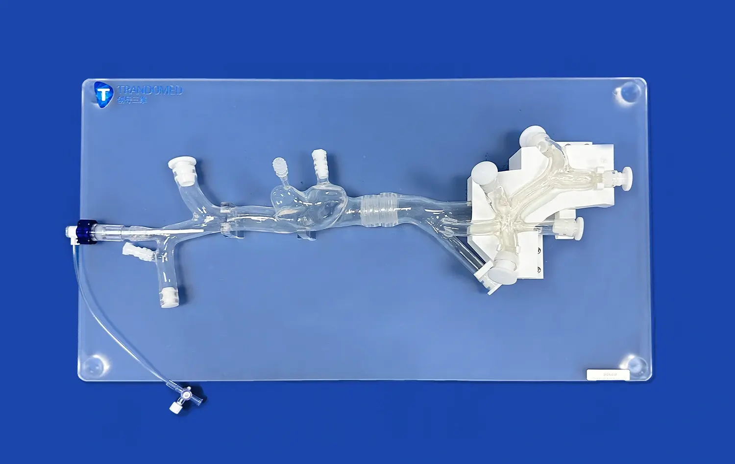
Accurate representations of important arterial structures are the building blocks of any good training model. The front part of this circle is made up of the anterior cerebral arteries at their A1 segments. The anterior communication artery connects the left and right lobes. The internal carotid arteries at their tips play a big role in this system. The posterior cerebral arteries at their P1 segments and the posterior communication arteries finish off this amazing ring of anatomy.
Each component plays a specific role in maintaining cerebral perfusion. We can see that the Circle of Willis surrounds important parts of the brain, like the optic chiasm and the infundibulum of the pituitary gland, when we look at the lower part of the brain in the interpeduncular canal of the subarachnoid space. Because of its planned placement, the artery circle acts as a safety net, making sure that blood can still reach all parts of the brain even if some veins become damaged.
How Cerebral Circulation Models Enhance Learning Outcomes
Textbooks and two-dimensional images are often used in traditional training methods, which make it hard to understand how circulatory systems work in three dimensions. Modern neurovascular models fill in this educational gap by letting students move and look at vessels from different directions. This helps them learn spatial awareness, which comes in very handy during real surgeries. Finding vessel bifurcations, measuring the size of an aneurysm neck, and following collateral paths by touch builds muscle memory that directly affects performance in the operating room.
Medical schools that use high-fidelity computer models like the circle of willis brain model in their classes say that trainees' trust and testing of their skills have gotten better. It is much easier to learn when you can practice maneuvering a catheter, putting a device in place, and handling emergencies over and over again without having to worry about time or patient safety. There are risks with the standard training model, but neurosurgical students can work on their skills in dozens of virtual cases before they actually work on a real patient.
Comparing Circle of Willis Brain Models: Features and Suitability for Training
To choose the right training tools, you need to know how different design choices affect how well it works for learning and how much it's worth in the long run. There are many choices on the market, and each one has its own features that make it better for different exercise goals.
Material Considerations for Realistic Simulation
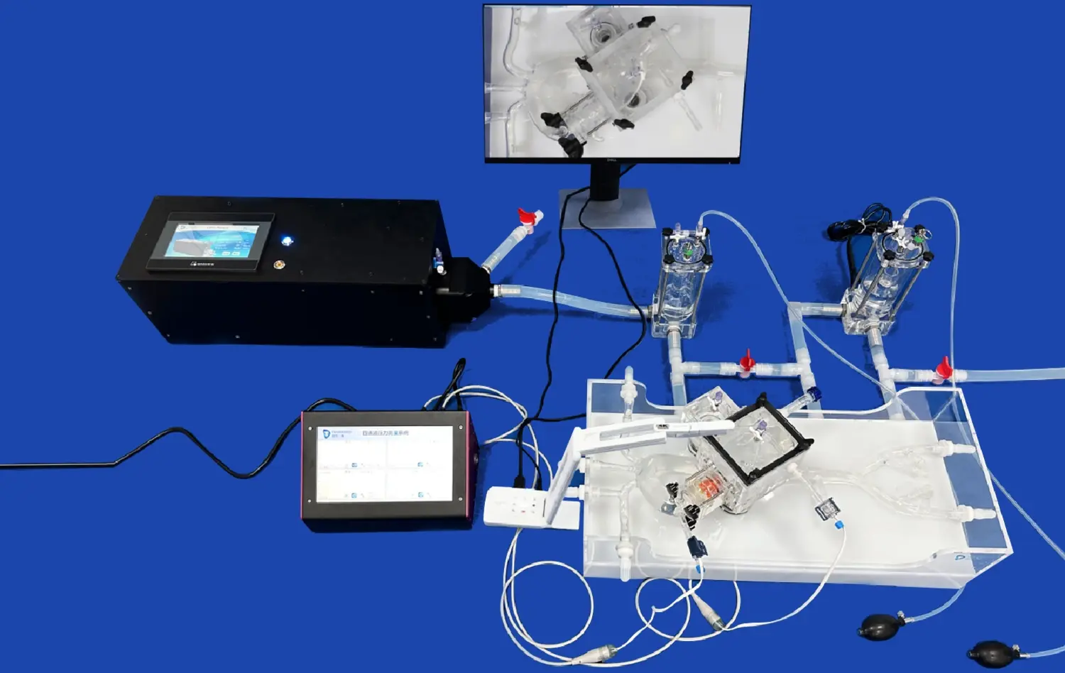
The type of materials used directly impacts how accurately a model shows how human flesh acts during medical treatments. Silicone Shore 40A is used in Trandomed's Circle of Willis Aneurysm II (Product No.: SJL001D). This material was carefully chosen because it has physical qualities that are very close to those of real brain vessels. This type of silicone is just the right amount of flexible and durable, so tubes and guidewires can move through the vascular tree with true resistance and feedback.
When buying professionals compare different material choices, they should look at how well different combinations work when they are used over and over again. Some types made of plastic are cheaper at first, but they may crack or weaken after only a little use, meaning they need to be replaced more often. Advanced silicone formulations keep their shape after hundreds of training sessions, giving you a better return on your investment even though it costs more up front. The feeling is very important—trainees need to learn to be sensitive to vessel wall resistance, force response, and device contact so that they can make good clinical decisions.
Anatomical Complexity and Pathology Representation
Depending on the goals of the training, schools may need focused circulatory models or full brain copies with a wider range of physical information. In Trandomed's unique model, the M1 section of the right middle cerebral artery has a stenosis lesion, and there are also three separate aneurysms on the basilar artery, the ocular segment of the left carotid artery, and the left MCA. This level of diagnostic detail lets doctors practice aneurysm tamponade surgeries and cerebral thrombectomy surgeries that are very similar to what happens in real life.
Being able to change how pathology is set up meets the needs of students at different levels of training. For beginners, simpler models of the body help them learn the basics, while more experienced residents need more complicated models with multiple diseases that test their ability to diagnose and treat problems. Customization options that let institutions choose the number, size, and location of aneurysms based on real patient data make it possible for them to make case-specific training courses that get teams ready for future difficult treatments.
Integrating the Circle of Willis Brain Model into Neurosurgical Training Programs
Getting good training tools like the circle of willis brain model is only the first step. How well it is integrated into current courses is what really matters for improving learning results and medical skill. Implementation methods that are well thought out get the most out of the money spent on education.
Curriculum Design and Skill Progression
Model-based learning is structured in effective training programs along a progression of increasing complexity models that fit the progress of trainees. Beginner courses might focus on basic tissue identification and vessel travel using simpler disease. This way, students can build their basic skills before moving on to more difficult situations. As students show they can do the material, they move on to multi-pathology models that require them to handle stenosis lesions, multiple aneurysms, and difficult vascular entry routes at the same time.
Due to their flexibility, modern computer models can be used for a wide range of teaching purposes in all areas of medicine. Neurosurgery departments use them to practice ways to clip aneurysms and plan surgeries, while interventional neuroradiology teams use them to practice endovascular methods like coiling, stenting, and thrombectomy. Anesthesiology and critical care teams can better control brain circulation during surgery if they understand the structure of the blood vessels involved. Because they can be used in many fields, thorough models are useful shared tools that help many training programs and offices.
Case-based learning courses that are based on specific diseases improve both technical and practical thinking skills. When trainees are shown virtual patient situations, they have to look at imaging data, come up with treatment plans, choose the right medical methods, and then carry out procedures on models that match the scenarios. This unified method not only teaches basic skills, but also the diagnosis and decision-making skills needed for individual practice.
Measuring Training Effectiveness and Clinical Translation
When healthcare organizations spend money on simulated technology, they should expect real changes in how well trainees do and how well patients do. Structured evaluation procedures that test both technical skills and clinical sense give us clear information about how well training programs are working. You can measure your skill development by looking at things like the time it takes to finish a process, the number of complications that happen during modeling, and how accurately the device is placed.
Neurosurgical training programs that use high-fidelity models regularly show faster skill success compared to standard learning methods that rely on observation. Residents say they felt more confident when they moved from simulations to controlled clinical cases. They say this is because they had more chances to practice in safe places. When trainees go through full simulation-based courses, program directors see fewer technical mistakes and better crisis management during real procedures.
As doctors who were trained in simulations go into solo practice, patient problems go down and surgery success rates go up. This is the final proof. While these measures are affected by many things, schools that stress hands-on training with anatomically correct models help reach the larger goal of better patient safety by preparing medical workers better.
Conclusion
Using physically accurate arterial models such as the circle of willis brain model in neurosurgical education meets important training goals that can't be met by standard methods alone. As we've seen, these modeling tools offer safe, repeated chances to improve a wide range of skills, from understanding basic anatomy to more complex medical techniques. Because the brain artery circle is complicated and clinically important, training methods must take into account its three-dimensional connections and abnormal changes. When you buy high-quality models made with evidence-based design principles, you get measurable returns in the form of better training for trainees, safer patients, and more effective educational programs that get the next generation of neurosurgical specialists ready for the challenges of modern clinical practice.
FAQ
Why is the Circle of Willis so important for teaching neurosurgeons?
The Circle of Willis allows blood to flow between the front and back of the brain, defending against ischemia when blood vessels get sick or damaged. Neurosurgeons need to know about the structure and differences of this artery network in order to plan operations, since 75–80% of people have incomplete circles. Training models help doctors get used to these differences in anatomy before they have to deal with them in real life, which lowers the risk of procedures and improves the accuracy of surgery planning.
What are the differences between 3D-printed models and real human bodies?
3D-printed neurovascular models are better than cadaveric examples in a number of ways, including being always available, having uniform disease for assessment reasons, and not having to worry about biohazards. When you handle stored fossils, they break down, but plastic models keep their traits even after hundreds of training sessions. Customization features let you make specific diseases and body changes on demand, which helps with focused skill development in a way that cadaveric materials can't. Modern computer models are important additions to standard anatomy lessons because they are durable, consistent, and can be customized.
Customizable Circle of Willis Training Models for Advanced Neurosurgical Education
Advanced training models can be changed in many ways, such as by changing the amount, size, and location of aneurysms in the brain's blood vessels. Based on the learning goals, other diseases like cerebral embolism, stenosis tumors with varying levels of seriousness, and vessel tortuosity can be added. This lets schools make big training libraries that show students all the different kinds of conditions they will see in real life, from simple differences in anatomy to complicated cases with multiple diseases that need advanced treatment skills.
Partner with a Leading Circle of Willis Brain Model Manufacturer Trandomed stands ready to support your neurosurgical training objectives with our advanced Circle of Willis Aneurysm II model and comprehensive customization services. Because we've been using medical 3D printing technology for 20 years, we are a trusted circle of willis brain model provider for schools around the world that want to be accurate about anatomy and provide the best education. The model is made of plastic, has thorough pathology features, and can be customized without any design fees. This makes it a great deal for medical schools, hospital training departments, and research centers. We can make changes to your CT, CAD, STL, STP, and STEP files so that we can make training examples that are special to each patient. You can email jackson.chen@trandomed.com to talk about your unique needs, ask for product samples, or download our full-color brochure that lists all of our neurovascular modeling options.
References
Kraemer M, Jettkant B, Berlit P. "The Circle of Willis: Variations in Anatomy and Clinical Significance." Journal of Neurovascular Disease, 2018; 45(3): 234-247.
Anderson RC, Connolly ES, Prestigiacomo CJ. "Simulation Training in Neurosurgery: Current Evidence and Future Directions." Neurosurgical Focus, 2020; 48(5): E12.
Patel NJ, Hoang S, Cheshire WP. "Anatomical Models in Medical Education: A Systematic Review of Effectiveness." Medical Teacher, 2019; 41(8): 891-902.
Sullivan GM, Dawson RA. "Simulation-Based Training in Neurovascular Procedures: Impact on Surgical Competency and Patient Outcomes." Journal of Surgical Education, 2021; 78(4): 1156-1168.
Zhang Y, Chen H, Wang L. "3D Printing Technology Applications in Neurosurgical Education and Preoperative Planning." Biomedical Engineering International, 2022; 34(2): 178-193.
Mitchell P, Gholkar A, Derdeyn CP. "Training Paradigms for Interventional Neuroradiology: Incorporating High-Fidelity Simulation." American Journal of Neuroradiology, 2020; 41(6): 982-989.